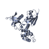
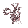
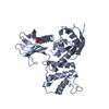
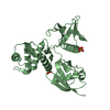
Entry Database : PDB / ID : 4rmaTitle Crystal structure of the FERM domain of human ezrin Ezrin Keywords / / / / Function / homology Function Domain/homology Component
/ / / / / / / / / / / / / / / / / / / / / / / / / / / / / / / / / / / / / / / / / / / / / / / / / / / / / / / / / / / / / / / / / / / / / / / / / / / / / / / / / / / / / / / / / / / / / / / / / / / / / / / / / / / / / / / / / / / / / / / / / / / / / / / / / / Biological species Homo sapiens (human)Method / / / Resolution : 1.75 Å Authors Phang, J.M. / Harrop, S.J. / Duff, A.P. / Wilk, K.E. / Curmi, P.M.G. Journal : Biochem. J. / Year : 2016Title : Structural characterization suggests models for monomeric and dimeric forms of full-length ezrin.Authors : Phang, J.M. / Harrop, S.J. / Duff, A.P. / Sokolova, A.V. / Crossett, B. / Walsh, J.C. / Beckham, S.A. / Nguyen, C.D. / Davies, R.B. / Glockner, C. / Bromley, E.H. / Wilk, K.E. / Curmi, P.M. History Deposition Oct 21, 2014 Deposition site / Processing site Revision 1.0 Dec 9, 2015 Provider / Type Revision 1.1 Sep 27, 2017 Group / Refinement description / Category / citation_author / softwareItem _citation.country / _citation.journal_abbrev ... _citation.country / _citation.journal_abbrev / _citation.journal_id_ASTM / _citation.journal_id_CSD / _citation.journal_id_ISSN / _citation.journal_volume / _citation.page_first / _citation.page_last / _citation.pdbx_database_id_DOI / _citation.pdbx_database_id_PubMed / _citation.title / _citation.year / _software.name Revision 1.2 Sep 20, 2023 Group Data collection / Database references ... Data collection / Database references / Derived calculations / Refinement description Category chem_comp_atom / chem_comp_bond ... chem_comp_atom / chem_comp_bond / database_2 / pdbx_initial_refinement_model / struct_site Item _database_2.pdbx_DOI / _database_2.pdbx_database_accession ... _database_2.pdbx_DOI / _database_2.pdbx_database_accession / _struct_site.pdbx_auth_asym_id / _struct_site.pdbx_auth_comp_id / _struct_site.pdbx_auth_seq_id
Show all Show less
 Movie
Movie Controller


 Open data
 Basic information
 Components
 Keywords
 Function and homology information
 Homo sapiens (human)
 X-RAY DIFFRACTION /
  Authors
 Citation
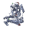
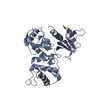
 Structure visualization
 Downloads & links
 Other downloads


 PDBj
PDBj


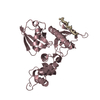
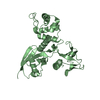
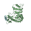
 Assembly
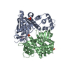


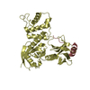
 Mass: 96.063 Da / Num. of mol.: 4 / Source method: obtained synthetically / Formula: SO4
 Mass: 18.015 Da / Num. of mol.: 450 / Source method: isolated from a natural source / Formula: H2O
 Sample preparation
 / Beamline: MX1 / Wavelength: 0.95369 Å
 Processing