 Movie
Movie Controller

[English] 日本語
 Yorodumi
- PDB-4qtu: Structure of S. cerevisiae Bud23-Trm112 complex involved in forma... -
+ Open data

- Basic information
| Entry | Database: PDB / ID: 4qtu | ||||||
|---|---|---|---|---|---|---|---|
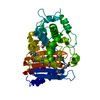
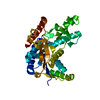
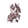
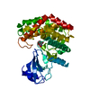
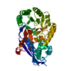
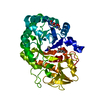
| Title | Structure of S. cerevisiae Bud23-Trm112 complex involved in formation of m7G1575 on 18S rRNA (SAM bound form) | ||||||
 Components Components |
| ||||||
 Keywords Keywords | TRANSFERASE / Class I / Methyltransferase | ||||||
| Function / homology |  Function and homology information Function and homology informationMethylation / positive regulation of termination of DNA-templated transcription / 18S rRNA (guanine1575-N7)-methyltransferase / eRF1 methyltransferase complex / Eukaryotic Translation Termination / tRNA (m2G10) methyltransferase complex / rRNA (guanine-N7)-methylation / tRNA methyltransferase activator activity / tRNA methyltransferase complex / rRNA (guanine) methyltransferase activity ...Methylation / positive regulation of termination of DNA-templated transcription / 18S rRNA (guanine1575-N7)-methyltransferase / eRF1 methyltransferase complex / Eukaryotic Translation Termination / tRNA (m2G10) methyltransferase complex / rRNA (guanine-N7)-methylation / tRNA methyltransferase activator activity / tRNA methyltransferase complex / rRNA (guanine) methyltransferase activity / tRNA wobble uridine modification / tRNA methylation / positive regulation of rRNA processing / maturation of LSU-rRNA / endonucleolytic cleavage in ITS1 to separate SSU-rRNA from 5.8S rRNA and LSU-rRNA from tricistronic rRNA transcript (SSU-rRNA, 5.8S rRNA, LSU-rRNA) / ribosomal small subunit export from nucleus / ribosomal large subunit biogenesis / maturation of SSU-rRNA / ribosomal small subunit biogenesis / ribosome / protein heterodimerization activity / nucleolus / nucleus / cytoplasm / cytosol Similarity search - Function | ||||||
| Biological species |  | ||||||
| Method |  X-RAY DIFFRACTION / SYNCHROTRON / MOLECULAR REPLACEMENT / Resolution: 2.124 Å X-RAY DIFFRACTION / SYNCHROTRON / MOLECULAR REPLACEMENT / Resolution: 2.124 Å | ||||||
 Authors Authors | Letoquart, J. / Huvelle, E. / Wacheul, L. / Bourgeois, G. / Zorbas, C. / Graille, M. / Heurgue-Hamard, V. / Lafontaine, D.L.J. | ||||||
 Citation Citation | Journal: Proc.Natl.Acad.Sci.USA / Year: 2014 Title: Structural and functional studies of Bud23-Trm112 reveal 18S rRNA N7-G1575 methylation occurs on late 40S precursor ribosomes. Authors: Letoquart, J. / Huvelle, E. / Wacheul, L. / Bourgeois, G. / Zorbas, C. / Graille, M. / Heurgue-Hamard, V. / Lafontaine, D.L. | ||||||
| History |
|
- Structure visualization
| Structure viewer | Molecule: MolmilJmol/JSmol |
|---|
- Downloads & links
-Download
| PDBx/mmCIF format | 4qtu.cif.gz | 144.6 KB | Display | PDBx/mmCIF format |
|---|---|---|---|---|
| PDB format | pdb4qtu.ent.gz | 112 KB | Display | PDB format |
| PDBx/mmJSON format | 4qtu.json.gz | Tree view | PDBx/mmJSON format | |
| Others |  Other downloads Other downloads |
-Validation report
| Arichive directory | https://data.pdbj.org/pub/pdb/validation_reports/qt/4qtuftp://data.pdbj.org/pub/pdb/validation_reports/qt/4qtu | HTTPS FTP |
|---|
-Related structure data
| Related structure data | 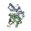 4qttSC  4qtm S: Starting model for refinement C: citing same article ( |
|---|---|
| Similar structure data |


-Links
 PDBj
PDBj
- Assembly
| Deposited unit | 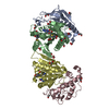
| ||||||||
|---|---|---|---|---|---|---|---|---|---|
| 1 | 
| ||||||||
| 2 | 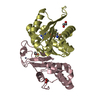
| ||||||||
| Unit cell |
|
-Components
-Protein , 2 types, 4 molecules ACBD
| #1: Protein | Mass: 15078.209 Da / Num. of mol.: 2 Source method: isolated from a genetically manipulated source Source: (gene. exp.)  #2: Protein | Mass: 23048.053 Da / Num. of mol.: 2 / Fragment: fragment residues 1-202 Source method: isolated from a genetically manipulated source Source: (gene. exp.) References: UniProt: P25627, Transferases; Transferring one-carbon groups; Methyltransferases |
|---|
-Non-polymers , 4 types, 196 molecules 


| #3: Chemical |  Mass: 65.409 Da / Num. of mol.: 2 / Source method: obtained synthetically / Formula: Zn Mass: 65.409 Da / Num. of mol.: 2 / Source method: obtained synthetically / Formula: Zn#4: Chemical | ChemComp-EDO /  Mass: 62.068 Da / Num. of mol.: 19 / Source method: obtained synthetically / Formula: C2H6O2 Mass: 62.068 Da / Num. of mol.: 19 / Source method: obtained synthetically / Formula: C2H6O2#5: Chemical |  Mass: 398.437 Da / Num. of mol.: 2 / Source method: obtained synthetically / Formula: C15H22N6O5S Mass: 398.437 Da / Num. of mol.: 2 / Source method: obtained synthetically / Formula: C15H22N6O5S#6: Water | ChemComp-HOH / | Mass: 18.015 Da / Num. of mol.: 173 / Source method: isolated from a natural source / Formula: H2O |
|---|
-Details
| Has protein modification | Y |
|---|
-Experimental details
-Experiment
| Experiment | Method: X-RAY DIFFRACTION / Number of used crystals: 1 |
|---|
- Sample preparation
| Crystal | Density Matthews: 2.26 Å3/Da / Density % sol: 45.54 % |
|---|---|
| Crystal grow | Temperature: 291 K / Method: vapor diffusion, hanging drop / pH: 7.5 Details: pH 7.5, VAPOR DIFFUSION, HANGING DROP, temperature 291K |
-Data collection
| Diffraction | Mean temperature: 100 K |
|---|---|
| Diffraction source | Source: SYNCHROTRON / Site: SOLEIL  / Beamline: PROXIMA 1 / Wavelength: 0.9801 Å / Beamline: PROXIMA 1 / Wavelength: 0.9801 Å |
| Detector | Type: PSI PILATUS 6M / Detector: PIXEL / Date: Dec 16, 2012 |
| Radiation | Monochromator: Channel cut cryogenically cooled monochromator crystal Protocol: SINGLE WAVELENGTH / Monochromatic (M) / Laue (L): M / Scattering type: x-ray |
| Radiation wavelength | Wavelength: 0.9801 Å / Relative weight: 1 |
| Reflection | Resolution: 2.124→50 Å / Num. obs: 38373 / % possible obs: 94.7 % / Observed criterion σ(F): 0 / Observed criterion σ(I): 0 / Biso Wilson estimate: 37.1 Å2 |
| Reflection shell | Resolution: 2.2→2.33 Å / % possible all: 94.7 |
- Processing
| Software |
| |||||||||||||||||||||||||||||||||||||||||||||||||||||||||||||||||||||||||||||||||||||||||||||||||||||||||
|---|---|---|---|---|---|---|---|---|---|---|---|---|---|---|---|---|---|---|---|---|---|---|---|---|---|---|---|---|---|---|---|---|---|---|---|---|---|---|---|---|---|---|---|---|---|---|---|---|---|---|---|---|---|---|---|---|---|---|---|---|---|---|---|---|---|---|---|---|---|---|---|---|---|---|---|---|---|---|---|---|---|---|---|---|---|---|---|---|---|---|---|---|---|---|---|---|---|---|---|---|---|---|---|---|---|---|
| Refinement | Method to determine structure: MOLECULAR REPLACEMENT Starting model: 4QTT Resolution: 2.124→38.307 Å / FOM work R set: 0.8226 / SU ML: 0.27 / σ(F): 1.99 / Phase error: 24.88 / Stereochemistry target values: ML
| |||||||||||||||||||||||||||||||||||||||||||||||||||||||||||||||||||||||||||||||||||||||||||||||||||||||||
| Solvent computation | Shrinkage radii: 0.9 Å / VDW probe radii: 1.11 Å / Solvent model: FLAT BULK SOLVENT MODEL | |||||||||||||||||||||||||||||||||||||||||||||||||||||||||||||||||||||||||||||||||||||||||||||||||||||||||
| Displacement parameters | Biso max: 101.94 Å2 / Biso mean: 44.88 Å2 / Biso min: 19.75 Å2 | |||||||||||||||||||||||||||||||||||||||||||||||||||||||||||||||||||||||||||||||||||||||||||||||||||||||||
| Refinement step | Cycle: LAST / Resolution: 2.124→38.307 Å
| |||||||||||||||||||||||||||||||||||||||||||||||||||||||||||||||||||||||||||||||||||||||||||||||||||||||||
| Refine LS restraints |
| |||||||||||||||||||||||||||||||||||||||||||||||||||||||||||||||||||||||||||||||||||||||||||||||||||||||||
| LS refinement shell | Refine-ID: X-RAY DIFFRACTION / Total num. of bins used: 14
|
