 Movie
Movie Controller


+ Open data

- Basic information
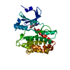
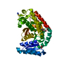
| Entry | Database: PDB / ID: 1x9y | ||||||
|---|---|---|---|---|---|---|---|
| Title | The prostaphopain B structure | ||||||
 Components Components | cysteine proteinase | ||||||
 Keywords Keywords | HYDROLASE / half-barrel / barrel-sandwich-hybrid | ||||||
| Function / homology |  Function and homology information Function and homology informationcysteine-type peptidase activity / Hydrolases; Acting on peptide bonds (peptidases); Cysteine endopeptidases / proteolysis / extracellular region Similarity search - Function | ||||||
| Biological species |   Staphylococcus aureus (bacteria) Staphylococcus aureus (bacteria) | ||||||
| Method |  X-RAY DIFFRACTION / MOLECULAR REPLACEMENT / Resolution: 2.5 Å X-RAY DIFFRACTION / MOLECULAR REPLACEMENT / Resolution: 2.5 Å | ||||||
 Authors Authors | Filipek, R. / Szczepanowski, R. / Sabat, A. / Potempa, J. / Bochtler, M. | ||||||
 Citation Citation | Journal: Biochemistry / Year: 2004 Title: Prostaphopain B structure: a comparison of proregion-mediated and staphostatin-mediated protease inhibition. Authors: Filipek, R. / Szczepanowski, R. / Sabat, A. / Potempa, J. / Bochtler, M. #1: Journal: J.Biol.Chem. / Year: 2003Title: The Staphostatin-staphopain complex: a forward binding inhibitor in complex with its target cysteine protease. Authors: Filipek, R. / Rzychon, M. / Oleksy, A. / Gruca, M. / Dubin, A. / Potempa, J. / Bochtler, M. | ||||||
| History |
|
- Structure visualization
| Structure viewer | Molecule: MolmilJmol/JSmol |
|---|
- Downloads & links
-Download
| PDBx/mmCIF format | 1x9y.cif.gz | 281.3 KB | Display | PDBx/mmCIF format |
|---|---|---|---|---|
| PDB format | pdb1x9y.ent.gz | 228.6 KB | Display | PDB format |
| PDBx/mmJSON format | 1x9y.json.gz | Tree view | PDBx/mmJSON format | |
| Others |  Other downloads Other downloads |
-Validation report
| Arichive directory | https://data.pdbj.org/pub/pdb/validation_reports/x9/1x9yftp://data.pdbj.org/pub/pdb/validation_reports/x9/1x9y | HTTPS FTP |
|---|
-Related structure data
| Related structure data | 1pxvS S: Starting model for refinement |
|---|---|
| Similar structure data |


-Links
 PDBj
PDBj
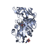
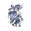
- Assembly
| Deposited unit | 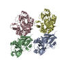
| ||||||||
|---|---|---|---|---|---|---|---|---|---|
| 1 | 
| ||||||||
| 2 | 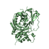
| ||||||||
| 3 | 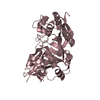
| ||||||||
| 4 | 
| ||||||||
| Unit cell |
| ||||||||
| Details | The protein is a monomer |
-Components
| #1: Protein | Mass: 41846.504 Da / Num. of mol.: 4 / Fragment: proenzyme (residues 37-393) Source method: isolated from a genetically manipulated source Source: (gene. exp.) Staphylococcus aureus (bacteria) / Gene: sspB / Plasmid: pGEX-5T / Species (production host): Escherichia coli / Production host: References: UniProt: Q70UQ8, UniProt: P0C1S6*PLUS, Hydrolases; Acting on peptide bonds (peptidases); Cysteine endopeptidases #2: Water | ChemComp-HOH / |  Mass: 18.015 Da / Num. of mol.: 243 / Source method: isolated from a natural source / Formula: H2O Mass: 18.015 Da / Num. of mol.: 243 / Source method: isolated from a natural source / Formula: H2O |
|---|
-Experimental details
-Experiment
| Experiment | Method: X-RAY DIFFRACTION / Number of used crystals: 1 |
|---|
- Sample preparation
| Crystal | Density Matthews: 2.47 Å3/Da / Density % sol: 49.8 % |
|---|---|
| Crystal grow | Temperature: 277.15 K / Method: vapor diffusion, sitting drop / pH: 6.5 Details: 20% PEG 4000, 50 mM Bis-Tris, 5 mM Tris, pH 6.5, VAPOR DIFFUSION, SITTING DROP, temperature 277.15K |
-Data collection
| Diffraction | Mean temperature: 100 K |
|---|---|
| Diffraction source | Source: ROTATING ANODE / Type: RIGAKU RU300 / Wavelength: 1.54 Å |
| Detector | Type: MARRESEARCH / Detector: IMAGE PLATE / Date: Oct 9, 2003 / Details: MSC Confocal MaxFlux mirrors |
| Radiation | Monochromator: MSC Confocal MaxFlux / Protocol: SINGLE WAVELENGTH / Monochromatic (M) / Laue (L): M / Scattering type: x-ray |
| Radiation wavelength | Wavelength: 1.54 Å / Relative weight: 1 |
| Reflection | Resolution: 2.5→20 Å / Num. all: 53566 / Num. obs: 53566 / % possible obs: 99.3 % / Observed criterion σ(F): 0 / Observed criterion σ(I): 0 / Redundancy: 2.7 % / Biso Wilson estimate: 46.9 Å2 / Rmerge(I) obs: 0.067 / Rsym value: 0.067 / Net I/σ(I): 17 |
| Reflection shell | Resolution: 2.5→2.54 Å / Redundancy: 2.6 % / Rmerge(I) obs: 0.339 / Mean I/σ(I) obs: 3 / Num. unique all: 2535 / Rsym value: 0.339 / % possible all: 95.6 |
- Processing
| Software |
| ||||||||||||||||||||||||||||||||||||
|---|---|---|---|---|---|---|---|---|---|---|---|---|---|---|---|---|---|---|---|---|---|---|---|---|---|---|---|---|---|---|---|---|---|---|---|---|---|
| Refinement | Method to determine structure: MOLECULAR REPLACEMENT Starting model: PDB entry 1PXV Resolution: 2.5→19.86 Å / Rfactor Rfree error: 0.005 / Data cutoff high absF: 2479432.46 / Data cutoff low absF: 0 / Isotropic thermal model: RESTRAINED / Cross valid method: THROUGHOUT / σ(F): 0 / Stereochemistry target values: Engh & Huber
| ||||||||||||||||||||||||||||||||||||
| Solvent computation | Solvent model: FLAT MODEL / Bsol: 24.0088 Å2 / ksol: 0.311264 e/Å3 | ||||||||||||||||||||||||||||||||||||
| Displacement parameters | Biso mean: 38 Å2
| ||||||||||||||||||||||||||||||||||||
| Refine analyze |
| ||||||||||||||||||||||||||||||||||||
| Refinement step | Cycle: LAST / Resolution: 2.5→19.86 Å
| ||||||||||||||||||||||||||||||||||||
| Refine LS restraints |
| ||||||||||||||||||||||||||||||||||||
| LS refinement shell | Resolution: 2.5→2.66 Å / Rfactor Rfree error: 0.017 / Total num. of bins used: 6
| ||||||||||||||||||||||||||||||||||||
| Xplor file |
|
