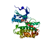
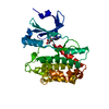
regulation of blood volume by renin-angiotensin / Metabolism of Angiotensinogen to Angiotensins / renin / juxtaglomerular apparatus development / mesonephros development / renin-angiotensin regulation of aldosterone production / response to cGMP / drinking behavior / response to immobilization stress / regulation of MAPK cascade ...regulation of blood volume by renin-angiotensin / Metabolism of Angiotensinogen to Angiotensins / renin / juxtaglomerular apparatus development / mesonephros development / renin-angiotensin regulation of aldosterone production / response to cGMP / drinking behavior / response to immobilization stress / regulation of MAPK cascade / amyloid-beta metabolic process / response to cAMP / cell maturation / angiotensin maturation / hormone-mediated signaling pathway / insulin-like growth factor receptor binding / male gonad development / cellular response to xenobiotic stimulus / regulation of blood pressure / apical part of cell / response to lipopolysaccharide / endopeptidase activity / aspartic-type endopeptidase activity / response to xenobiotic stimulus / signaling receptor binding / : / membrane Similarity search - Function
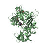
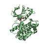
Renin-like domain / Aspartic peptidase, N-terminal / A1 Propeptide / Eukaryotic aspartyl protease / Aspartic peptidase A1 family / Peptidase family A1 domain / Peptidase family A1 domain profile. / Cathepsin D, subunit A; domain 1 / Acid Proteases / Aspartic peptidase, active site ...Renin-like domain / Aspartic peptidase, N-terminal / A1 Propeptide / Eukaryotic aspartyl protease / Aspartic peptidase A1 family / Peptidase family A1 domain / Peptidase family A1 domain profile. / Cathepsin D, subunit A; domain 1 / Acid Proteases / Aspartic peptidase, active site / Eukaryotic and viral aspartyl proteases active site. / Aspartic peptidase domain superfamily / Beta Barrel / Mainly Beta Similarity search - Domain/homology
Resolution: 3.2→50.21 Å / Cor.coef. Fo:Fc: 0.899 / Cor.coef. Fo:Fc free: 0.897 / SU B: 30.333 / SU ML: 0.457 / Cross valid method: THROUGHOUT / ESU R: 0.764 / ESU R Free: 0.422 / Details: HYDROGENS HAVE BEEN ADDED IN THE RIDING POSITIONS
Rfactor
Num. reflection
% reflection
Selection details
Rfree
0.29723
703
5.2 %
RANDOM
Rwork
0.26308
-
-
-
obs
0.26499
12878
99.9 %
-
Solvent computation
Ion probe radii: 0.8 Å / Shrinkage radii: 0.8 Å / VDW probe radii: 1.2 Å
 Movie
Movie Controller


 Open data
 Basic information
 Components
 Keywords
 Function and homology information
 X-RAY DIFFRACTION /
  Authors
 United Kingdom, 2items
  Citation
 Structure visualization
 Downloads & links
 Other downloads
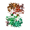


 PDBj
PDBj
 Assembly
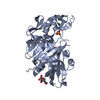
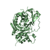
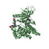
 Homo sapiens (human) / References: UniProt: P06281, renin
 Type: D-saccharide, beta linking / Mass: 221.208 Da / Num. of mol.: 1
 Sample preparation
 Processing