- PDB-4qma: Crystal Structure of a Putative Cysteine Dioxygnase From Ralstoni... -
+
Open data
ID or keywords:
Loading...
-
Basic information
Entry
Database: PDB / ID: 4qma
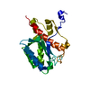
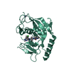
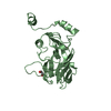
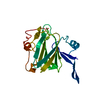
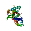
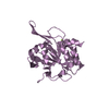
Title
Crystal Structure of a Putative Cysteine Dioxygnase From Ralstonia eutropha: An Alternative Modeling of 2GM6 from JCSG Target 361076
Components
Cysteine dioxygenase type I
Keywords
OXIDOREDUCTASE / PUTATIVE "Gln-type" CYSTEINE DIOXYGENASE / JOINT CENTER FOR STRUCTURAL GENOMICS
Function / homology
Function and homology information
oxidoreductase activity, acting on single donors with incorporation of molecular oxygen, incorporation of two atoms of oxygen / ferrous iron binding Similarity search - Function
ATP synthase delta/epsilon subunit, C-terminal domain / Cysteine dioxygenase type I / Cysteine dioxygenase type I / RmlC-like cupin domain superfamily / Jelly Rolls / Single alpha-helices involved in coiled-coils or other helix-helix interfaces / RmlC-like jelly roll fold / Jelly Rolls / Up-down Bundle / Sandwich ...ATP synthase delta/epsilon subunit, C-terminal domain / Cysteine dioxygenase type I / Cysteine dioxygenase type I / RmlC-like cupin domain superfamily / Jelly Rolls / Single alpha-helices involved in coiled-coils or other helix-helix interfaces / RmlC-like jelly roll fold / Jelly Rolls / Up-down Bundle / Sandwich / Mainly Beta / Mainly Alpha Similarity search - Domain/homology
Resolution: 1.65→44.748 Å / SU ML: 0.21 / σ(F): 0.1 / Phase error: 26.22 / Stereochemistry target values: ML Details: 1. HYDROGENS HAVE BEEN ADDED IN THE RIDING POSITIONS. 2. A MET-INHIBITION PROTOCOL WAS USED FOR SELENOMETHIONINE INCORPORATION DURING PROTEIN EXPRESSION. THE OCCUPANCY OF THE SE ATOMS IN THE ...Details: 1. HYDROGENS HAVE BEEN ADDED IN THE RIDING POSITIONS. 2. A MET-INHIBITION PROTOCOL WAS USED FOR SELENOMETHIONINE INCORPORATION DURING PROTEIN EXPRESSION. THE OCCUPANCY OF THE SE ATOMS IN THE MSE RESIDUES WAS REDUCED TO 0.75 FOR THE REDUCED SCATTERING POWER DUE TO PARTIAL S-MET INCORPORATION. 3. FE MODELED BASED ON ANOMALOUS DIFFERENCE MAP, GEOMETRY, PROTEIN ENVIRONMENT, AND EXCITATION SCAN. 4. SO4 AND EDO MODELED BASED ON CYRSTALLIZATION CONDITIONS.
Rfactor
Num. reflection
% reflection
Selection details
Rfree
0.2
2217
5 %
Same as ReCDO, random 5% to 1.65 Ang
Rwork
0.1741
-
-
-
obs
0.1754
42041
99.87 %
-
all
-
82002
-
-
Solvent computation
Shrinkage radii: 0.9 Å / VDW probe radii: 1.11 Å / Solvent model: FLAT BULK SOLVENT MODEL
Refinement step
Cycle: LAST / Resolution: 1.65→44.748 Å
Protein
Nucleic acid
Ligand
Solvent
Total
Num. atoms
1499
0
49
209
1757
Refine LS restraints
Refine-ID
Type
Dev ideal
Number
X-RAY DIFFRACTION
f_bond_d
0.014
1637
X-RAY DIFFRACTION
f_angle_d
1.213
2213
X-RAY DIFFRACTION
f_dihedral_angle_d
13.641
602
X-RAY DIFFRACTION
f_chiral_restr
0.052
227
X-RAY DIFFRACTION
f_plane_restr
0.006
293
LS refinement shell
Resolution (Å)
Rfactor Rfree
Num. reflection Rfree
Rfactor Rwork
Num. reflection Rwork
Refine-ID
% reflection obs (%)
1.65-1.6694
0.4201
127
0.3627
2697
X-RAY DIFFRACTION
100
1.6694-1.6898
0.3563
121
0.3602
2727
X-RAY DIFFRACTION
100
1.6898-1.7112
0.3465
127
0.3628
2683
X-RAY DIFFRACTION
100
1.7112-1.7337
0.4053
151
0.3396
2690
X-RAY DIFFRACTION
100
1.7337-1.7574
0.3304
148
0.3285
2676
X-RAY DIFFRACTION
100
1.7574-1.7825
0.3238
131
0.3249
2714
X-RAY DIFFRACTION
100
1.7825-1.8091
0.2909
137
0.3138
2648
X-RAY DIFFRACTION
100
1.8091-1.8374
0.287
131
0.3144
2757
X-RAY DIFFRACTION
100
1.8374-1.8675
0.304
127
0.2887
2632
X-RAY DIFFRACTION
100
1.8675-1.8997
0.3292
166
0.2755
2694
X-RAY DIFFRACTION
100
1.8997-1.9343
0.2425
156
0.245
2622
X-RAY DIFFRACTION
100
1.9343-1.9715
0.2754
139
0.238
2716
X-RAY DIFFRACTION
100
1.9715-2.0117
0.2612
143
0.2286
2669
X-RAY DIFFRACTION
100
2.0117-2.0555
0.2361
137
0.2159
2698
X-RAY DIFFRACTION
100
2.0555-2.1033
0.2621
117
0.2198
2720
X-RAY DIFFRACTION
100
2.1033-2.1559
0.2193
157
0.1979
2646
X-RAY DIFFRACTION
100
2.1559-2.2142
0.2215
165
0.179
2669
X-RAY DIFFRACTION
100
2.2142-2.2793
0.1748
158
0.1706
2709
X-RAY DIFFRACTION
100
2.2793-2.3529
0.2255
161
0.1645
2648
X-RAY DIFFRACTION
100
2.3529-2.437
0.1911
140
0.1658
2684
X-RAY DIFFRACTION
100
2.437-2.5345
0.1819
129
0.158
2687
X-RAY DIFFRACTION
100
2.5345-2.6499
0.1966
136
0.1539
2689
X-RAY DIFFRACTION
100
2.6499-2.7896
0.2039
146
0.162
2701
X-RAY DIFFRACTION
100
2.7896-2.9643
0.2296
163
0.1761
2693
X-RAY DIFFRACTION
100
2.9643-3.1931
0.179
145
0.1725
2662
X-RAY DIFFRACTION
100
3.1931-3.5144
0.2129
141
0.1596
2689
X-RAY DIFFRACTION
100
3.5144-4.0226
0.1557
134
0.1486
2692
X-RAY DIFFRACTION
100
4.0226-5.067
0.135
134
0.1224
2699
X-RAY DIFFRACTION
100
5.067-44.7646
0.1962
136
0.1658
2688
X-RAY DIFFRACTION
100
Refinement TLS params.
Method: refined / Origin x: 36.5971 Å / Origin y: 21.0005 Å / Origin z: 37.2235 Å
11
12
13
21
22
23
31
32
33
T
0.3632 Å2
0.0757 Å2
-0.0615 Å2
-
0.2898 Å2
-0.0215 Å2
-
-
0.269 Å2
L
0.9058 °2
0.4243 °2
-0.4857 °2
-
1.4719 °2
-0.638 °2
-
-
3.055 °2
S
-0.1417 Å °
0.0201 Å °
0.1114 Å °
-0.196 Å °
0.0637 Å °
0.1759 Å °
-0.203 Å °
-0.3025 Å °
0.0594 Å °
Refinement TLS group
Selection details: chain 'A' and resid 11 through 202
+
About Yorodumi
-
News
-
Feb 9, 2022. New format data for meta-information of EMDB entries
New format data for meta-information of EMDB entries
Version 3 of the EMDB header file is now the official format.
The previous official version 1.9 will be removed from the archive.
In the structure databanks used in Yorodumi, some data are registered as the other names, "COVID-19 virus" and "2019-nCoV". Here are the details of the virus and the list of structure data.
Jan 31, 2019. EMDB accession codes are about to change! (news from PDBe EMDB page)
EMDB accession codes are about to change! (news from PDBe EMDB page)
The allocation of 4 digits for EMDB accession codes will soon come to an end. Whilst these codes will remain in use, new EMDB accession codes will include an additional digit and will expand incrementally as the available range of codes is exhausted. The current 4-digit format prefixed with “EMD-” (i.e. EMD-XXXX) will advance to a 5-digit format (i.e. EMD-XXXXX), and so on. It is currently estimated that the 4-digit codes will be depleted around Spring 2019, at which point the 5-digit format will come into force.
The EM Navigator/Yorodumi systems omit the EMD- prefix.
Related info.:Q: What is EMD? / ID/Accession-code notation in Yorodumi/EM Navigator
Yorodumi is a browser for structure data from EMDB, PDB, SASBDB, etc.
This page is also the successor to EM Navigator detail page, and also detail information page/front-end page for Omokage search.
The word "yorodu" (or yorozu) is an old Japanese word meaning "ten thousand". "mi" (miru) is to see.
Related info.:EMDB / PDB / SASBDB / Comparison of 3 databanks / Yorodumi Search / Aug 31, 2016. New EM Navigator & Yorodumi / Yorodumi Papers / Jmol/JSmol / Function and homology information / Changes in new EM Navigator and Yorodumi
 Movie
Movie Controller

 Yorodumi
 Open data
 Basic information
 Components
 Keywords
 Function and homology information
 Ralstonia eutropha JMP134 (bacteria)
 X-RAY DIFFRACTION /
  Authors
 Citation
 Structure visualization
 Downloads & links
 Other downloads
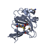


 PDBj
PDBj


 Assembly
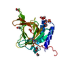


 Mass: 55.845 Da / Num. of mol.: 1 / Source method: obtained synthetically / Formula: Fe
 Mass: 31.999 Da / Num. of mol.: 1 / Source method: obtained synthetically / Formula: O2
 Mass: 96.063 Da / Num. of mol.: 2 / Source method: obtained synthetically / Formula: SO4
 Mass: 62.068 Da / Num. of mol.: 9 / Source method: obtained synthetically / Formula: C2H6O2
 Sample preparation
 Processing