- PDB-3uss: Crystal structure of Cysteine dioxygenase from Pseudomonas aeruginosa -
+
Open data
ID or keywords:
Loading...
-
Basic information
Entry
Database: PDB / ID: 3uss
Title
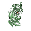
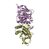
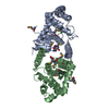
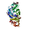
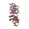
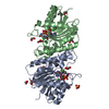
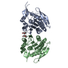
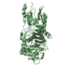
Crystal structure of Cysteine dioxygenase from Pseudomonas aeruginosa
Components
Putative uncharacterized protein
Keywords
OXIDOREDUCTASE / cupin / three histidine / non-heme iron / cysteine catabolism
Function / homology
Function and homology information
3-mercaptopropionate dioxygenase / oxidoreductase activity, acting on single donors with incorporation of molecular oxygen, incorporation of two atoms of oxygen / ferrous iron binding / iron ion binding Similarity search - Function
ATP synthase delta/epsilon subunit, C-terminal domain / Cysteine dioxygenase type I / Cysteine dioxygenase type I / RmlC-like cupin domain superfamily / Jelly Rolls / Single alpha-helices involved in coiled-coils or other helix-helix interfaces / RmlC-like jelly roll fold / Jelly Rolls / Up-down Bundle / Sandwich ...ATP synthase delta/epsilon subunit, C-terminal domain / Cysteine dioxygenase type I / Cysteine dioxygenase type I / RmlC-like cupin domain superfamily / Jelly Rolls / Single alpha-helices involved in coiled-coils or other helix-helix interfaces / RmlC-like jelly roll fold / Jelly Rolls / Up-down Bundle / Sandwich / Mainly Beta / Mainly Alpha Similarity search - Domain/homology
In the structure databanks used in Yorodumi, some data are registered as the other names, "COVID-19 virus" and "2019-nCoV". Here are the details of the virus and the list of structure data.
Jan 31, 2019. EMDB accession codes are about to change! (news from PDBe EMDB page)
EMDB accession codes are about to change! (news from PDBe EMDB page)
The allocation of 4 digits for EMDB accession codes will soon come to an end. Whilst these codes will remain in use, new EMDB accession codes will include an additional digit and will expand incrementally as the available range of codes is exhausted. The current 4-digit format prefixed with “EMD-” (i.e. EMD-XXXX) will advance to a 5-digit format (i.e. EMD-XXXXX), and so on. It is currently estimated that the 4-digit codes will be depleted around Spring 2019, at which point the 5-digit format will come into force.
The EM Navigator/Yorodumi systems omit the EMD- prefix.
Related info.:Q: What is EMD? / ID/Accession-code notation in Yorodumi/EM Navigator
Yorodumi is a browser for structure data from EMDB, PDB, SASBDB, etc.
This page is also the successor to EM Navigator detail page, and also detail information page/front-end page for Omokage search.
The word "yorodu" (or yorozu) is an old Japanese word meaning "ten thousand". "mi" (miru) is to see.
Related info.:EMDB / PDB / SASBDB / Comparison of 3 databanks / Yorodumi Search / Aug 31, 2016. New EM Navigator & Yorodumi / Yorodumi Papers / Jmol/JSmol / Function and homology information / Changes in new EM Navigator and Yorodumi
 Movie
Movie Controller

 Yorodumi
 Open data
 Basic information
 Components
 Keywords
 Function and homology information
 Pseudomonas aeruginosa (bacteria)
 X-RAY DIFFRACTION /
  Authors
 Citation
 Structure visualization
 Downloads & links
 Other downloads


 PDBj
PDBj


 Assembly


 Mass: 55.845 Da / Num. of mol.: 2 / Source method: obtained synthetically / Formula: Fe
 Mass: 96.063 Da / Num. of mol.: 9 / Source method: obtained synthetically / Formula: SO4
 Mass: 22.990 Da / Num. of mol.: 4 / Source method: obtained synthetically / Formula: Na
 Mass: 35.453 Da / Num. of mol.: 1 / Source method: obtained synthetically / Formula: Cl
 Sample preparation
 / Beamline: MX1 / Wavelength: 0.9537 Å
 Processing