 Movie
Movie Controller

[English] 日本語
 Yorodumi
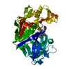
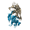
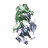
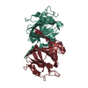
- PDB-1epz: CRYSTAL STRUCTURE OF DTDP-6-DEOXY-D-XYLO-4-HEXULOASE 3,5-EPIMERAS... -
+ Open data

- Basic information
| Entry | Database: PDB / ID: 1epz | ||||||
|---|---|---|---|---|---|---|---|
| Title | CRYSTAL STRUCTURE OF DTDP-6-DEOXY-D-XYLO-4-HEXULOASE 3,5-EPIMERASE FROM METHANOBACTERIUM THERMOAUTOTROPHICUM WITH BOUND LIGAND. | ||||||
 Components Components | DTDP-6-DEOXY-D-XYLO-4-HEXULOSE 3,5-EPIMERASE | ||||||
 Keywords Keywords | ISOMERASE / racemase / dTDP-4-dehydrorhamnose epimerase | ||||||
| Function / homology |  Function and homology information Function and homology informationdTDP-4-dehydrorhamnose 3,5-epimerase / dTDP-4-dehydrorhamnose 3,5-epimerase activity / dTDP-rhamnose biosynthetic process / polysaccharide biosynthetic process / cytosol Similarity search - Function | ||||||
| Biological species |   Methanothermobacter thermautotrophicus (archaea) Methanothermobacter thermautotrophicus (archaea) | ||||||
| Method |  X-RAY DIFFRACTION / Resolution: 1.75 Å X-RAY DIFFRACTION / Resolution: 1.75 Å | ||||||
 Authors Authors | Christendat, D. / Saridakis, V. / Bochkarev, A. / Pai, E.F. / Arrowsmith, C. / Edwards, A.M. | ||||||
 Citation Citation | Journal: J.Biol.Chem. / Year: 2000 Title: Crystal structure of dTDP-4-keto-6-deoxy-D-hexulose 3,5-epimerase from Methanobacterium thermoautotrophicum complexed with dTDP. Authors: Christendat, D. / Saridakis, V. / Dharamsi, A. / Bochkarev, A. / Pai, E.F. / Arrowsmith, C.H. / Edwards, A.M. | ||||||
| History |
|
- Structure visualization
| Structure viewer | Molecule: MolmilJmol/JSmol |
|---|
- Downloads & links
-Download
| PDBx/mmCIF format | 1epz.cif.gz | 53.4 KB | Display | PDBx/mmCIF format |
|---|---|---|---|---|
| PDB format | pdb1epz.ent.gz | 37.8 KB | Display | PDB format |
| PDBx/mmJSON format | 1epz.json.gz | Tree view | PDBx/mmJSON format | |
| Others |  Other downloads Other downloads |
-Validation report
| Arichive directory | https://data.pdbj.org/pub/pdb/validation_reports/ep/1epzftp://data.pdbj.org/pub/pdb/validation_reports/ep/1epz | HTTPS FTP |
|---|
-Related structure data


-Links
 PDBj
PDBj- Assembly
| Deposited unit | 
| ||||||||
|---|---|---|---|---|---|---|---|---|---|
| 1 | 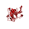
| ||||||||
| Unit cell |
| ||||||||
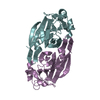
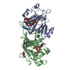
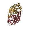
| Details | The biological assembly is a dimer constructed from Chain A and one symmetry related molecule. |
-Components
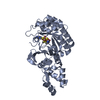
| #1: Protein | Mass: 21702.332 Da / Num. of mol.: 1 Source method: isolated from a genetically manipulated source Source: (gene. exp.) Methanothermobacter thermautotrophicus (archaea)Plasmid: PET15B / Production host:  References: UniProt: O27818, dTDP-4-dehydrorhamnose 3,5-epimerase |
|---|---|
| #2: Chemical | ChemComp-TYD /   Mass: 402.188 Da / Num. of mol.: 1 / Source method: obtained synthetically / Formula: C10H16N2O11P2 Mass: 402.188 Da / Num. of mol.: 1 / Source method: obtained synthetically / Formula: C10H16N2O11P2 |
| #3: Water | ChemComp-HOH /  Mass: 18.015 Da / Num. of mol.: 119 / Source method: isolated from a natural source / Formula: H2O Mass: 18.015 Da / Num. of mol.: 119 / Source method: isolated from a natural source / Formula: H2O |
-Experimental details
-Experiment
| Experiment | Method: X-RAY DIFFRACTION / Number of used crystals: 1 |
|---|
- Sample preparation
| Crystal | Density Matthews: 2.14 Å3/Da / Density % sol: 42.6 % | |||||||||||||||
|---|---|---|---|---|---|---|---|---|---|---|---|---|---|---|---|---|
| Crystal grow | Temperature: 293 K / Method: vapor diffusion, hanging drop / pH: 4.6 Details: 10% polyethylene glycol 4K, 0.1 M Sodium Acetate, pH 4.6, VAPOR DIFFUSION, HANGING DROP, temperature 20K | |||||||||||||||
| Crystal | *PLUS Density % sol: 46 % | |||||||||||||||
| Crystal grow | *PLUS Temperature: 22 ℃ | |||||||||||||||
| Components of the solutions | *PLUS
|
-Data collection
| Diffraction | Mean temperature: 100 K |
|---|---|
| Diffraction source | Source: ROTATING ANODE / Type: RIGAKU RU200 / Wavelength: 1.5418 |
| Detector | Type: MARRESEARCH / Detector: IMAGE PLATE / Date: Mar 28, 2000 |
| Radiation | Protocol: SINGLE WAVELENGTH / Monochromatic (M) / Laue (L): M / Scattering type: x-ray |
| Radiation wavelength | Wavelength: 1.5418 Å / Relative weight: 1 |
| Reflection | Resolution: 1.75→25 Å / Num. all: 63707 / Num. obs: 17671 / % possible obs: 95 % / Observed criterion σ(F): 4862 / Observed criterion σ(I): 206 / Redundancy: 4 % / Biso Wilson estimate: 14.2 Å2 / Rmerge(I) obs: 0.052 / Net I/σ(I): 24 |
| Reflection shell | Resolution: 1.75→1.81 Å / Redundancy: 4 % / Rmerge(I) obs: 0.321 / Num. unique all: 1708 / % possible all: 92 |
| Reflection | *PLUS % possible obs: 95.2 % / Rmerge(I) obs: 0.041 |
| Reflection shell | *PLUS % possible obs: 92 % / Rmerge(I) obs: 0.295 / Mean I/σ(I) obs: 4 |
- Processing
| Software |
| ||||||||||||||||||||||||||||||||||||||||||||||||||||||||||||||||||||||||||||||||
|---|---|---|---|---|---|---|---|---|---|---|---|---|---|---|---|---|---|---|---|---|---|---|---|---|---|---|---|---|---|---|---|---|---|---|---|---|---|---|---|---|---|---|---|---|---|---|---|---|---|---|---|---|---|---|---|---|---|---|---|---|---|---|---|---|---|---|---|---|---|---|---|---|---|---|---|---|---|---|---|---|---|
| Refinement | Resolution: 1.75→9.99 Å / Rfactor Rfree error: 0.005 / Data cutoff high absF: 456278.04 / Data cutoff low absF: 0 / Isotropic thermal model: RESTRAINED / Cross valid method: THROUGHOUT / σ(F): 0 / Stereochemistry target values: CNS 0.9 / Details: Simulated Annealing
| ||||||||||||||||||||||||||||||||||||||||||||||||||||||||||||||||||||||||||||||||
| Solvent computation | Solvent model: FLAT MODEL / Bsol: 56.66 Å2 / ksol: 0.536 e/Å3 | ||||||||||||||||||||||||||||||||||||||||||||||||||||||||||||||||||||||||||||||||
| Displacement parameters | Biso mean: 18.5 Å2
| ||||||||||||||||||||||||||||||||||||||||||||||||||||||||||||||||||||||||||||||||
| Refine analyze |
| ||||||||||||||||||||||||||||||||||||||||||||||||||||||||||||||||||||||||||||||||
| Refinement step | Cycle: LAST / Resolution: 1.75→9.99 Å
| ||||||||||||||||||||||||||||||||||||||||||||||||||||||||||||||||||||||||||||||||
| Refine LS restraints |
| ||||||||||||||||||||||||||||||||||||||||||||||||||||||||||||||||||||||||||||||||
| LS refinement shell | Resolution: 1.75→1.86 Å / Rfactor Rfree error: 0.019 / Total num. of bins used: 6
| ||||||||||||||||||||||||||||||||||||||||||||||||||||||||||||||||||||||||||||||||
| Xplor file |
| ||||||||||||||||||||||||||||||||||||||||||||||||||||||||||||||||||||||||||||||||
| Software | *PLUS Name: CNS / Version: 0.9 / Classification: refinement | ||||||||||||||||||||||||||||||||||||||||||||||||||||||||||||||||||||||||||||||||
| Refine LS restraints | *PLUS
|
