 Movie
Movie Controller

[English] 日本語
 Yorodumi
- PDB-1wlt: Crystal Structure of dTDP-4-dehydrorhamnose 3,5-epimerase homolog... -
+ Open data

- Basic information
| Entry | Database: PDB / ID: 1wlt | ||||||
|---|---|---|---|---|---|---|---|
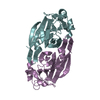
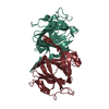
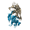
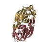
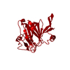
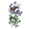
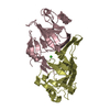
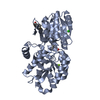
| Title | Crystal Structure of dTDP-4-dehydrorhamnose 3,5-epimerase homologue from Sulfolobus tokodaii | ||||||
 Components Components | 176aa long hypothetical dTDP-4-dehydrorhamnose 3,5-epimerase | ||||||
 Keywords Keywords | ISOMERASE / jelly roll-like topology / flattened barrel | ||||||
| Function / homology |  Function and homology information Function and homology informationdTDP-4-dehydrorhamnose 3,5-epimerase / dTDP-4-dehydrorhamnose 3,5-epimerase activity / dTDP-rhamnose biosynthetic process / polysaccharide biosynthetic process / cytosol Similarity search - Function | ||||||
| Biological species |   Sulfolobus tokodaii (archaea) Sulfolobus tokodaii (archaea) | ||||||
| Method |  X-RAY DIFFRACTION / SYNCHROTRON / MOLECULAR REPLACEMENT / Resolution: 1.9 Å X-RAY DIFFRACTION / SYNCHROTRON / MOLECULAR REPLACEMENT / Resolution: 1.9 Å | ||||||
 Authors Authors | Watanabe, N. / Sakai, N. / Zilian, Z. / Kawarabayasi, Y. / Tanaka, I. | ||||||
 Citation Citation | Journal: to be published Title: Crystal Structure of dTDP-4-dehydrorhamnose 3,5-epimerase homologue Authors: Watanabe, N. / Sakai, N. / Zilian, Z. / Kawarabayasi, Y. / Tanaka, I. | ||||||
| History |
|
- Structure visualization
| Structure viewer | Molecule: MolmilJmol/JSmol |
|---|
- Downloads & links
-Download
| PDBx/mmCIF format | 1wlt.cif.gz | 91.1 KB | Display | PDBx/mmCIF format |
|---|---|---|---|---|
| PDB format | pdb1wlt.ent.gz | 69.1 KB | Display | PDB format |
| PDBx/mmJSON format | 1wlt.json.gz | Tree view | PDBx/mmJSON format | |
| Others |  Other downloads Other downloads |
-Validation report
| Arichive directory | https://data.pdbj.org/pub/pdb/validation_reports/wl/1wltftp://data.pdbj.org/pub/pdb/validation_reports/wl/1wlt | HTTPS FTP |
|---|
-Related structure data
| Related structure data |  1dzrS S: Starting model for refinement |
|---|---|
| Similar structure data |

-Links
 PDBj
PDBj- Assembly
| Deposited unit | 
| ||||||||
|---|---|---|---|---|---|---|---|---|---|
| 1 |
| ||||||||
| Unit cell |
| ||||||||
| Details | The biological assembly is a dimer in the asymmetric unit. |
-Components
| #1: Protein | Mass: 22677.107 Da / Num. of mol.: 2 Source method: isolated from a genetically manipulated source Source: (gene. exp.) Sulfolobus tokodaii (archaea) / Plasmid: pET23q / Production host:  References: UniProt: Q96Z62, dTDP-4-dehydrorhamnose 3,5-epimerase #2: Water | ChemComp-HOH / |  Mass: 18.015 Da / Num. of mol.: 272 / Source method: isolated from a natural source / Formula: H2O Mass: 18.015 Da / Num. of mol.: 272 / Source method: isolated from a natural source / Formula: H2OHas protein modification | Y | |
|---|
-Experimental details
-Experiment
| Experiment | Method: X-RAY DIFFRACTION / Number of used crystals: 1 |
|---|
- Sample preparation
| Crystal | Density Matthews: 2.5 Å3/Da / Density % sol: 49.4 % |
|---|---|
| Crystal grow | Temperature: 293 K / Method: vapor diffusion, sitting drop / pH: 6.5 Details: 15% PEG 3350, 0.1M Sodium cacodylate buffer, pH 6.5, VAPOR DIFFUSION, SITTING DROP, temperature 293K |
-Data collection
| Diffraction | Mean temperature: 95 K |
|---|---|
| Diffraction source | Source: SYNCHROTRON / Site: Photon Factory  / Beamline: BL-6A / Wavelength: 1 Å / Beamline: BL-6A / Wavelength: 1 Å |
| Detector | Type: ADSC QUANTUM 4 / Detector: CCD / Date: Mar 2, 2004 / Details: MONOCHROMETER |
| Radiation | Monochromator: Si 111 / Protocol: SINGLE WAVELENGTH / Monochromatic (M) / Laue (L): M / Scattering type: x-ray |
| Radiation wavelength | Wavelength: 1 Å / Relative weight: 1 |
| Reflection | Resolution: 1.89→50 Å / Num. all: 35811 / Num. obs: 35811 / % possible obs: 99.9 % / Observed criterion σ(I): -3 / Redundancy: 6.6 % / Biso Wilson estimate: 24.5 Å2 / Rsym value: 0.06 / Net I/σ(I): 15.5 |
| Reflection shell | Resolution: 1.89→1.96 Å / Redundancy: 6.3 % / Mean I/σ(I) obs: 6.43 / Num. unique all: 3486 / Rsym value: 0.379 / % possible all: 99.7 |
- Processing
| Software |
| ||||||||||||||||||||||||||||||||||||||||||||||||||||||||||||||||||||||||||||||||||||||||||||||||||||
|---|---|---|---|---|---|---|---|---|---|---|---|---|---|---|---|---|---|---|---|---|---|---|---|---|---|---|---|---|---|---|---|---|---|---|---|---|---|---|---|---|---|---|---|---|---|---|---|---|---|---|---|---|---|---|---|---|---|---|---|---|---|---|---|---|---|---|---|---|---|---|---|---|---|---|---|---|---|---|---|---|---|---|---|---|---|---|---|---|---|---|---|---|---|---|---|---|---|---|---|---|---|
| Refinement | Method to determine structure: MOLECULAR REPLACEMENT Starting model: PDB ENTRY 1DZR Resolution: 1.9→20 Å / Cor.coef. Fo:Fc: 0.951 / Cor.coef. Fo:Fc free: 0.927 / SU B: 3.349 / SU ML: 0.1 / Cross valid method: THROUGHOUT / ESU R: 0.15 / ESU R Free: 0.147 / Stereochemistry target values: MAXIMUM LIKELIHOOD / Details: HYDROGENS HAVE BEEN ADDED IN THE RIDING POSITIONS
| ||||||||||||||||||||||||||||||||||||||||||||||||||||||||||||||||||||||||||||||||||||||||||||||||||||
| Solvent computation | Ion probe radii: 0.8 Å / Shrinkage radii: 0.8 Å / VDW probe radii: 1.4 Å / Solvent model: BABINET MODEL WITH MASK | ||||||||||||||||||||||||||||||||||||||||||||||||||||||||||||||||||||||||||||||||||||||||||||||||||||
| Displacement parameters | Biso mean: 26.019 Å2
| ||||||||||||||||||||||||||||||||||||||||||||||||||||||||||||||||||||||||||||||||||||||||||||||||||||
| Refinement step | Cycle: LAST / Resolution: 1.9→20 Å
| ||||||||||||||||||||||||||||||||||||||||||||||||||||||||||||||||||||||||||||||||||||||||||||||||||||
| Refine LS restraints |
| ||||||||||||||||||||||||||||||||||||||||||||||||||||||||||||||||||||||||||||||||||||||||||||||||||||
| LS refinement shell | Resolution: 1.9→1.949 Å / Total num. of bins used: 20
|
