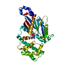
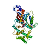
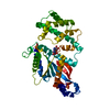
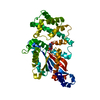
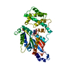
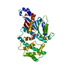
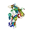
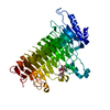
Entry Database : PDB / ID : 4panTitle A conserved phenylalanine as relay between the 5 helix and the GDP binding region of heterotrimeric G protein Guanine nucleotide-binding protein G(i) subunit alpha-1 Keywords / / / / / Function / homology Function Domain/homology Component
/ / / / / / / / / / / / / / / / / / / / / / / / / / / / / / / / / / / / / / / / / / / / / / / / / / / / / / / / / / / / / / / / / / / / / / / / Biological species Rattus norvegicus (Norway rat)Method / / Resolution : 2.4 Å Authors Kaya, A.I. / Lokits, A.D. / Gilbert, J. / Iverson, T.M. / Meiler, J. / Hamm, H.E. Funding support Organization Grant number Country National Institutes of Health/National Eye Institute (NIH/NEI) RO1 EY006062 National Institutes of Health/National Institute of General Medical Sciences (NIH/NIGMS) RO1 GM095633 National Institutes of Health/National Institute of General Medical Sciences (NIH/NIGMS) RO1 GM080403 National Institutes of Health/National Institute of Mental Health (NIH/NIMH) RO1 MH090192 National Institutes of Health/National Institute of Mental Health (NIH/NIMH) RO1 MH090192 National Institutes of Health/National Institute of General Medical Sciences (NIH/NIGMS) RO1 GM099842 National Institutes of Health/National Institute of Diabetes and Digestive and Kidney Disease (NIH/NIDDK) RO1 DK09737 National Institutes of Health/National Center for Research Resources (NIH/NCRR) S10 RR026915 National Science Foundation (NSF, United States) 0742762 National Science Foundation (NSF, United States) 1305874
Journal : J.Biol.Chem. / Year : 2014Title : A Conserved Phenylalanine as a Relay between the alpha 5 Helix and the GDP Binding Region of Heterotrimeric Gi Protein alpha Subunit.Authors : Kaya, A.I. / Lokits, A.D. / Gilbert, J.A. / Iverson, T.M. / Meiler, J. / Hamm, H.E. History Deposition Apr 9, 2014 Deposition site / Processing site Revision 1.0 Jul 30, 2014 Provider / Type Revision 1.1 Oct 1, 2014 Group Revision 1.2 Sep 13, 2017 Group Author supporting evidence / Database references ... Author supporting evidence / Database references / Derived calculations / Other / Source and taxonomy Category citation / entity_src_gen ... citation / entity_src_gen / pdbx_audit_support / pdbx_database_status / pdbx_struct_oper_list Item _citation.journal_id_CSD / _entity_src_gen.pdbx_alt_source_flag ... _citation.journal_id_CSD / _entity_src_gen.pdbx_alt_source_flag / _pdbx_audit_support.funding_organization / _pdbx_database_status.pdb_format_compatible / _pdbx_struct_oper_list.symmetry_operation Revision 1.3 Nov 27, 2019 Group / Category / Item Revision 1.4 Mar 30, 2022 Group / Database references / Refinement descriptionCategory / pdbx_audit_support / refine_histItem / _database_2.pdbx_database_accession / _pdbx_audit_support.funding_organizationRevision 1.5 Dec 27, 2023 Group / Category / chem_comp_bond
Show all Show less
 Movie
Movie Controller

 Yorodumi
 Open data
 Basic information
 Components
 Keywords
 Function and homology information
 X-RAY DIFFRACTION /
  Authors
 United States, 10items
  Citation
 Structure visualization
 Downloads & links
 Other downloads
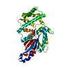


 PDBj
PDBj


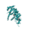
 Assembly
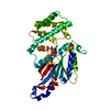


 Type: RNA linking / Mass: 443.201 Da / Num. of mol.: 1 / Source method: obtained synthetically / Formula: C10H15N5O11P2 / Comment: GDP, energy-carrying molecule*YM
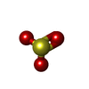
 Mass: 80.063 Da / Num. of mol.: 1 / Source method: obtained synthetically / Formula: SO3
 Mass: 35.453 Da / Num. of mol.: 1 / Source method: obtained synthetically / Formula: Cl
 Mass: 18.015 Da / Num. of mol.: 88 / Source method: isolated from a natural source / Formula: H2O
 Sample preparation
 Processing