 Movie
Movie Controller

[English] 日本語
 Yorodumi
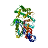
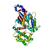
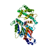
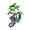
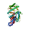
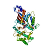
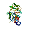
- PDB-1gdd: TERTIARY AND QUATERNARY STRUCTURAL CHANGES IN GIA1 INDUCED BY GTP... -
+ Open data

- Basic information
| Entry | Database: PDB / ID: 1gdd | ||||||
|---|---|---|---|---|---|---|---|
| Title | TERTIARY AND QUATERNARY STRUCTURAL CHANGES IN GIA1 INDUCED BY GTP HYDROLYSIS | ||||||
 Components Components | GI ALPHA 1 | ||||||
 Keywords Keywords | SIGNAL TRANSDUCTION PROTEIN / GTP-ASE | ||||||
| Function / homology |  Function and homology information Function and homology informationadenylate cyclase regulator activity / Extra-nuclear estrogen signaling / Adenylate cyclase inhibitory pathway / negative regulation of synaptic transmission / Adrenaline,noradrenaline inhibits insulin secretion / ADP signalling through P2Y purinoceptor 12 / GTPase activating protein binding / G alpha (i) signalling events / neurotransmitter receptor localization to postsynaptic specialization membrane / adenylate cyclase inhibitor activity ...adenylate cyclase regulator activity / Extra-nuclear estrogen signaling / Adenylate cyclase inhibitory pathway / negative regulation of synaptic transmission / Adrenaline,noradrenaline inhibits insulin secretion / ADP signalling through P2Y purinoceptor 12 / GTPase activating protein binding / G alpha (i) signalling events / neurotransmitter receptor localization to postsynaptic specialization membrane / adenylate cyclase inhibitor activity / positive regulation of protein localization to cell cortex / T cell migration / D2 dopamine receptor binding / adenylate cyclase-inhibiting serotonin receptor signaling pathway / G protein-coupled serotonin receptor binding / cellular response to forskolin / regulation of mitotic spindle organization / chemokine-mediated signaling pathway / response to prostaglandin E / positive regulation of cholesterol biosynthetic process / negative regulation of insulin secretion / G protein-coupled receptor binding / centriolar satellite / G-protein beta/gamma-subunit complex binding / adenylate cyclase-modulating G protein-coupled receptor signaling pathway / adenylate cyclase-inhibiting G protein-coupled receptor signaling pathway / GDP binding / heterotrimeric G-protein complex / G protein activity / midbody / cell cortex / Hydrolases; Acting on acid anhydrides; Acting on GTP to facilitate cellular and subcellular movement / postsynapse / ciliary basal body / G protein-coupled receptor signaling pathway / cell division / GTPase activity / centrosome / GTP binding / nucleolus / glutamatergic synapse / magnesium ion binding / Golgi apparatus / protein-containing complex / nucleoplasm / plasma membrane / cytoplasm / cytosol Similarity search - Function | ||||||
| Biological species |  | ||||||
| Method |  X-RAY DIFFRACTION / Resolution: 2.2 Å X-RAY DIFFRACTION / Resolution: 2.2 Å | ||||||
 Authors Authors | Mixon, M.B. / Sprang, S.R. | ||||||
 Citation Citation | Journal: Science / Year: 1995 Title: Tertiary and quaternary structural changes in Gi alpha 1 induced by GTP hydrolysis. Authors: Mixon, M.B. / Lee, E. / Coleman, D.E. / Berghuis, A.M. / Gilman, A.G. / Sprang, S.R. #1: Journal: J.Mol.Biol. / Year: 1994Title: Crystallization and Preliminary Crystallographic Studies of Gi Alpha 1 and Mutants of Gi Alpha 1 in the GTP and Gdp-Bound States Authors: Coleman, D.E. / Lee, E. / Mixon, M.B. / Linder, M.E. / Berghuis, A.M. / Gilman, A.G. / Sprang, S.R. #2: Journal: Science / Year: 1994Title: Structures of Active Conformations of Gi Alpha 1 and the Mechanism of GTP Hydrolysis Authors: Coleman, D.E. / Berghuis, A.M. / Lee, E. / Linder, M.E. / Gilman, A.G. / Sprang, S.R. | ||||||
| History |
|
- Structure visualization
| Structure viewer | Molecule: MolmilJmol/JSmol |
|---|
- Downloads & links
-Download
| PDBx/mmCIF format | 1gdd.cif.gz | 79.5 KB | Display | PDBx/mmCIF format |
|---|---|---|---|---|
| PDB format | pdb1gdd.ent.gz | 59.1 KB | Display | PDB format |
| PDBx/mmJSON format | 1gdd.json.gz | Tree view | PDBx/mmJSON format | |
| Others |  Other downloads Other downloads |
-Validation report
| Arichive directory | https://data.pdbj.org/pub/pdb/validation_reports/gd/1gddftp://data.pdbj.org/pub/pdb/validation_reports/gd/1gdd | HTTPS FTP |
|---|
-Related structure data
| Similar structure data |
|---|

-Links
 PDBj
PDBj


- Assembly
| Deposited unit | 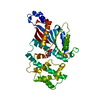
| ||||||||
|---|---|---|---|---|---|---|---|---|---|
| 1 |
| ||||||||
| Unit cell |
|
-Components
| #1: Protein | Mass: 40267.836 Da / Num. of mol.: 1 Source method: isolated from a genetically manipulated source Details: INACTIVE FORM / Source: (gene. exp.)  |
|---|---|
| #2: Chemical | ChemComp-SO4 /   Mass: 96.063 Da / Num. of mol.: 1 / Source method: obtained synthetically / Formula: SO4 Mass: 96.063 Da / Num. of mol.: 1 / Source method: obtained synthetically / Formula: SO4 |
| #3: Chemical | ChemComp-GDP /   Type: RNA linking / Mass: 443.201 Da / Num. of mol.: 1 / Source method: obtained synthetically / Formula: C10H15N5O11P2 / Comment: GDP, energy-carrying molecule*YM Type: RNA linking / Mass: 443.201 Da / Num. of mol.: 1 / Source method: obtained synthetically / Formula: C10H15N5O11P2 / Comment: GDP, energy-carrying molecule*YM |
| #4: Water | ChemComp-HOH /  Mass: 18.015 Da / Num. of mol.: 70 / Source method: isolated from a natural source / Formula: H2O Mass: 18.015 Da / Num. of mol.: 70 / Source method: isolated from a natural source / Formula: H2O |
-Experimental details
-Experiment
| Experiment | Method: X-RAY DIFFRACTION |
|---|
- Sample preparation
| Crystal | Density Matthews: 3.15 Å3/Da / Density % sol: 60.97 % | ||||||||||||||||||||||||||||||||||||||||||||||||||||||||||||
|---|---|---|---|---|---|---|---|---|---|---|---|---|---|---|---|---|---|---|---|---|---|---|---|---|---|---|---|---|---|---|---|---|---|---|---|---|---|---|---|---|---|---|---|---|---|---|---|---|---|---|---|---|---|---|---|---|---|---|---|---|---|
| Crystal grow | *PLUS pH: 8 / Method: vapor diffusion | ||||||||||||||||||||||||||||||||||||||||||||||||||||||||||||
| Components of the solutions | *PLUS
|
-Data collection
| Diffraction | Mean temperature: 110 K |
|---|---|
| Diffraction source | Wavelength: 1.5418 |
| Detector | Type: RIGAKU RAXIS IIC / Detector: IMAGE PLATE / Date: Apr 12, 1995 |
| Radiation | Monochromatic (M) / Laue (L): M / Scattering type: x-ray |
| Radiation wavelength | Wavelength: 1.5418 Å / Relative weight: 1 |
| Reflection | Resolution: 2.2→20 Å / Num. obs: 23024 / % possible obs: 95 % / Observed criterion σ(I): 1 / Redundancy: 1.2 % / Rmerge(I) obs: 0.052 |
| Reflection | *PLUS Rmerge(I) obs: 0.052 |
| Reflection shell | *PLUS Highest resolution: 2.2 Å / Lowest resolution: 2.3 Å / % possible obs: 70.6 % / Rmerge(I) obs: 0.412 / Mean I/σ(I) obs: 2.4 |
- Processing
| Software |
| ||||||||||||||||||||||||||||||||||||||||||||||||||||||||||||
|---|---|---|---|---|---|---|---|---|---|---|---|---|---|---|---|---|---|---|---|---|---|---|---|---|---|---|---|---|---|---|---|---|---|---|---|---|---|---|---|---|---|---|---|---|---|---|---|---|---|---|---|---|---|---|---|---|---|---|---|---|---|
| Refinement | Resolution: 2.2→6 Å / σ(F): 1
| ||||||||||||||||||||||||||||||||||||||||||||||||||||||||||||
| Displacement parameters | Biso mean: 38 Å2 | ||||||||||||||||||||||||||||||||||||||||||||||||||||||||||||
| Refinement step | Cycle: LAST / Resolution: 2.2→6 Å
| ||||||||||||||||||||||||||||||||||||||||||||||||||||||||||||
| Refine LS restraints |
| ||||||||||||||||||||||||||||||||||||||||||||||||||||||||||||
| Software | *PLUS Name: X-PLOR / Classification: refinement | ||||||||||||||||||||||||||||||||||||||||||||||||||||||||||||
| Refinement | *PLUS | ||||||||||||||||||||||||||||||||||||||||||||||||||||||||||||
| Solvent computation | *PLUS | ||||||||||||||||||||||||||||||||||||||||||||||||||||||||||||
| Displacement parameters | *PLUS | ||||||||||||||||||||||||||||||||||||||||||||||||||||||||||||
| Refine LS restraints | *PLUS
|
