 Movie
Movie Controller

[English] 日本語
 Yorodumi
- PDB-4a4m: Crystal structure of the light-activated constitutively active N2... -
+ Open data

- Basic information
| Entry | Database: PDB / ID: 4a4m | |||||||||
|---|---|---|---|---|---|---|---|---|---|---|
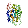
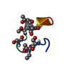
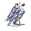
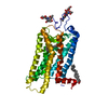
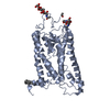
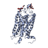
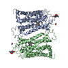
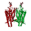
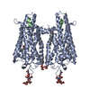
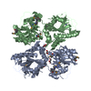
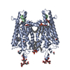
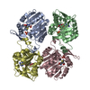
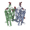
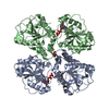
| Title | Crystal structure of the light-activated constitutively active N2C, M257Y,D282C rhodopsin mutant in complex with a peptide resembling the C-terminus of the Galpha-protein subunit (GaCT) | |||||||||
 Components Components |
| |||||||||
 Keywords Keywords | SIGNALING PROTEIN / G-PROTEIN / G-PROTEIN-COUPLED RECEPTORS / SIGNAL TANSDUCTION / VISUAL SYSTEM / METARHODOPSIN-II | |||||||||
| Function / homology |  Function and homology information Function and homology informationnegative regulation of cyclic-nucleotide phosphodiesterase activity / Adenylate cyclase inhibitory pathway / sensory perception of bitter taste / Opsins / VxPx cargo-targeting to cilium / rod bipolar cell differentiation / sperm head plasma membrane / absorption of visible light / opsin binding / The canonical retinoid cycle in rods (twilight vision) ...negative regulation of cyclic-nucleotide phosphodiesterase activity / Adenylate cyclase inhibitory pathway / sensory perception of bitter taste / Opsins / VxPx cargo-targeting to cilium / rod bipolar cell differentiation / sperm head plasma membrane / absorption of visible light / opsin binding / The canonical retinoid cycle in rods (twilight vision) / G protein-coupled opsin signaling pathway / Sensory perception of sweet, bitter, and umami (glutamate) taste / 11-cis retinal binding / Synthesis, secretion, and inactivation of Glucagon-like Peptide-1 (GLP-1) / podosome assembly / G protein-coupled photoreceptor activity / photoreceptor inner segment membrane / sensory perception of umami taste / cellular response to light stimulus / rod photoreceptor outer segment / detection of light stimulus involved in visual perception / sensory perception of sweet taste / G protein-coupled receptor complex / Inactivation, recovery and regulation of the phototransduction cascade / thermotaxis / Activation of the phototransduction cascade / outer membrane / detection of temperature stimulus involved in thermoception / response to light intensity / photoreceptor cell maintenance / arrestin family protein binding / ADP signalling through P2Y purinoceptor 12 / Extra-nuclear estrogen signaling / photoreceptor outer segment membrane / G alpha (i) signalling events / acyl binding / response to light stimulus / phototransduction, visible light / phototransduction / G-protein alpha-subunit binding / photoreceptor outer segment / photoreceptor inner segment / visual perception / guanyl-nucleotide exchange factor activity / G protein-coupled receptor binding / microtubule cytoskeleton organization / adenylate cyclase-modulating G protein-coupled receptor signaling pathway / G-protein beta/gamma-subunit complex binding / cell-cell junction / photoreceptor disc membrane / GDP binding / sperm midpiece / heterotrimeric G-protein complex / gene expression / G protein-coupled receptor signaling pathway / Golgi membrane / GTPase activity / protein kinase binding / GTP binding / zinc ion binding / metal ion binding / identical protein binding / membrane / plasma membrane / cytoplasm Similarity search - Function | |||||||||
| Biological species |  | |||||||||
| Method |  X-RAY DIFFRACTION / SYNCHROTRON / MOLECULAR REPLACEMENT / Resolution: 3.3 Å X-RAY DIFFRACTION / SYNCHROTRON / MOLECULAR REPLACEMENT / Resolution: 3.3 Å | |||||||||
 Authors Authors | Deupi, X. / Edwards, P. / Singhal, A. / Nickle, B. / Oprian, D.D. / Schertler, G.F.X. / Standfuss, J. | |||||||||
 Citation Citation | Journal: Proc.Natl.Acad.Sci.USA / Year: 2012 Title: Stabilized G Protein Binding Site in the Structure of Constitutively Active Metarhodopsin-II. Authors: Deupi, X. / Edwards, P. / Singhal, A. / Nickle, B. / Oprian, D. / Schertler, G. / Standfuss, J. #1: Journal: J.Mol.Biol. / Year: 2007Title: Crystal Structure of a Thermally Stable Rhodopsin Mutant. Authors: Standfuss, J. / Xie, G. / Edwards, P.C. / Burghammer, M. / Oprian, D.D. / Schertler, G.F.X. #2: Journal: Nature / Year: 2011Title: The Structural Basis of Agonist-Induced Activation in Constitutively Active Rhodopsin. Authors: Standfuss, J. / Edwards, P.C. / D'Antona, A. / Fransen, M. / Xie, G. / Oprian, D.D. / Schertler, G.F.X. | |||||||||
| History |
|
- Structure visualization
| Structure viewer | Molecule: MolmilJmol/JSmol |
|---|
- Downloads & links
-Download
| PDBx/mmCIF format | 4a4m.cif.gz | 86.6 KB | Display | PDBx/mmCIF format |
|---|---|---|---|---|
| PDB format | pdb4a4m.ent.gz | 63.6 KB | Display | PDB format |
| PDBx/mmJSON format | 4a4m.json.gz | Tree view | PDBx/mmJSON format | |
| Others |  Other downloads Other downloads |
-Validation report
| Arichive directory | https://data.pdbj.org/pub/pdb/validation_reports/a4/4a4mftp://data.pdbj.org/pub/pdb/validation_reports/a4/4a4m | HTTPS FTP |
|---|
-Related structure data
| Related structure data |  2x72S S: Starting model for refinement |
|---|---|
| Similar structure data |


-Links
 PDBj
PDBj


- Assembly
| Deposited unit | 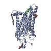
| ||||||||
|---|---|---|---|---|---|---|---|---|---|
| 1 | 
| ||||||||
| Unit cell |
|
-Components
-Protein / Protein/peptide , 2 types, 2 molecules AB
| #1: Protein | Mass: 39066.562 Da / Num. of mol.: 1 / Mutation: YES Source method: isolated from a genetically manipulated source Source: (gene. exp.)  HOMO SAPIENS (human) / References: UniProt: P02699 HOMO SAPIENS (human) / References: UniProt: P02699 |
|---|---|
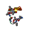
| #2: Protein/peptide | Mass: 1265.499 Da / Num. of mol.: 1 / Fragment: RESIDUES 344-354 / Mutation: YES / Source method: obtained synthetically / Source: (synth.) |
-Sugars , 2 types, 2 molecules 
| #3: Polysaccharide | 2-acetamido-2-deoxy-beta-D-glucopyranose-(1-4)-2-acetamido-2-deoxy-beta-D-glucopyranose Source method: isolated from a genetically manipulated source |
|---|---|
| #6: Sugar | ChemComp-BOG /  Type: D-saccharide / Mass: 292.369 Da / Num. of mol.: 1 Type: D-saccharide / Mass: 292.369 Da / Num. of mol.: 1Source method: isolated from a genetically manipulated source Formula: C14H28O6 / Comment: detergent*YM |
-Non-polymers , 4 types, 12 molecules 


| #4: Chemical | ChemComp-RET /  Mass: 284.436 Da / Num. of mol.: 1 / Source method: obtained synthetically / Formula: C20H28O Mass: 284.436 Da / Num. of mol.: 1 / Source method: obtained synthetically / Formula: C20H28O |
|---|---|
| #5: Chemical | ChemComp-ACT /  Mass: 59.044 Da / Num. of mol.: 1 / Source method: obtained synthetically / Formula: C2H3O2 Mass: 59.044 Da / Num. of mol.: 1 / Source method: obtained synthetically / Formula: C2H3O2 |
| #7: Chemical | ChemComp-PLM /  Mass: 256.424 Da / Num. of mol.: 1 / Source method: obtained synthetically / Formula: C16H32O2 Mass: 256.424 Da / Num. of mol.: 1 / Source method: obtained synthetically / Formula: C16H32O2 |
| #8: Water | ChemComp-HOH / Mass: 18.015 Da / Num. of mol.: 9 / Source method: isolated from a natural source / Formula: H2O |
-Details
| Compound details | ENGINEERED RESIDUE IN CHAIN A, ASN 2 TO CYS ENGINEERED RESIDUE IN CHAIN A, MET 257 TO TYR ...ENGINEERED |
|---|---|
| Has protein modification | Y |
-Experimental details
-Experiment
| Experiment | Method: X-RAY DIFFRACTION / Number of used crystals: 3 |
|---|
- Sample preparation
| Crystal | Density Matthews: 7.96 Å3/Da / Density % sol: 84.43 % / Description: NONE |
|---|---|
| Crystal grow | pH: 4.5 Details: 3.0-3.4 M AMMONIUM SULPHATE, 100 MM SODIUM ACETATE PH 4.5 |
-Data collection
| Diffraction | Mean temperature: 100 K |
|---|---|
| Diffraction source | Source: SYNCHROTRON / Site: SLS  / Beamline: X06SA / Wavelength: 1 / Beamline: X06SA / Wavelength: 1 |
| Detector | Type: DECTRIS PILATUS 6M / Detector: PIXEL |
| Radiation | Protocol: SINGLE WAVELENGTH / Monochromatic (M) / Laue (L): M / Scattering type: x-ray |
| Radiation wavelength | Wavelength: 1 Å / Relative weight: 1 |
| Reflection | Resolution: 3.3→45 Å / Num. obs: 17649 / % possible obs: 94.8 % / Observed criterion σ(I): -3 / Redundancy: 5.7 % / Biso Wilson estimate: 75.56 Å2 / Rmerge(I) obs: 0.18 / Net I/σ(I): 8.5 |
| Reflection shell | Resolution: 3.3→3.48 Å / Redundancy: 2.9 % / Rmerge(I) obs: 0.66 / Mean I/σ(I) obs: 1.8 / % possible all: 65.4 |
- Processing
| Software |
| |||||||||||||||||||||||||||||||||||||||||||||||||
|---|---|---|---|---|---|---|---|---|---|---|---|---|---|---|---|---|---|---|---|---|---|---|---|---|---|---|---|---|---|---|---|---|---|---|---|---|---|---|---|---|---|---|---|---|---|---|---|---|---|---|
| Refinement | Method to determine structure: MOLECULAR REPLACEMENT Starting model: PDB ENTRY 2X72 Resolution: 3.3→45.77 Å / SU ML: 0.42 / σ(F): 1.35 / Phase error: 26.86 / Stereochemistry target values: ML
| |||||||||||||||||||||||||||||||||||||||||||||||||
| Solvent computation | Shrinkage radii: 0.9 Å / VDW probe radii: 1.11 Å / Solvent model: FLAT BULK SOLVENT MODEL / Bsol: 40.896 Å2 / ksol: 0.279 e/Å3 | |||||||||||||||||||||||||||||||||||||||||||||||||
| Displacement parameters |
| |||||||||||||||||||||||||||||||||||||||||||||||||
| Refinement step | Cycle: LAST / Resolution: 3.3→45.77 Å
| |||||||||||||||||||||||||||||||||||||||||||||||||
| Refine LS restraints |
| |||||||||||||||||||||||||||||||||||||||||||||||||
| LS refinement shell |
|
