 Movie
Movie Controller

[English] 日本語
 Yorodumi
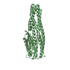
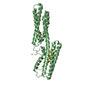
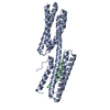
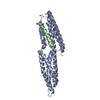
- PDB-3tj6: human vinculin head domain (Vh1, residues 1-258) in complex with ... -
+ Open data

- Basic information
| Entry | Database: PDB / ID: 3tj6 | ||||||
|---|---|---|---|---|---|---|---|
| Title | human vinculin head domain (Vh1, residues 1-258) in complex with the vinculin binding site of the surface cell antigen 4 (sca4-VBS-C; residues 812-835) from Rickettsia rickettsii | ||||||
 Components Components |
| ||||||
 Keywords Keywords | protein binding/toxin / cytoskeleton / epidemic typhus / sca4 / spotted fever / ALPHA-HELIX BUNDLE DOMAIN / PROTEIN-PROTEIN INTERACTIONS / CELL ADHESION / CYTOSOL / FOCAL ADHESION / protein binding-toxin complex | ||||||
| Function / homology |  Function and homology information Function and homology informationregulation of protein localization to adherens junction / outer dense plaque of desmosome / inner dense plaque of desmosome / podosome ring / terminal web / cell-substrate junction / epithelial cell-cell adhesion / zonula adherens / fascia adherens / dystroglycan binding ...regulation of protein localization to adherens junction / outer dense plaque of desmosome / inner dense plaque of desmosome / podosome ring / terminal web / cell-substrate junction / epithelial cell-cell adhesion / zonula adherens / fascia adherens / dystroglycan binding / alpha-catenin binding / cell-cell contact zone / apical junction assembly / costamere / regulation of establishment of endothelial barrier / axon extension / protein localization to cell surface / adherens junction assembly / lamellipodium assembly / regulation of focal adhesion assembly / maintenance of blood-brain barrier / brush border / Smooth Muscle Contraction / negative regulation of cell migration / Turbulent (oscillatory, disturbed) flow shear stress activates signaling by PIEZO1 and integrins in endothelial cells / morphogenesis of an epithelium / cell projection / cell-matrix adhesion / adherens junction / Signaling by high-kinase activity BRAF mutants / MAP2K and MAPK activation / beta-catenin binding / sarcolemma / platelet aggregation / specific granule lumen / Signaling by RAF1 mutants / Signaling by moderate kinase activity BRAF mutants / Paradoxical activation of RAF signaling by kinase inactive BRAF / Signaling downstream of RAS mutants / cell-cell junction / Signaling by BRAF and RAF1 fusions / Signaling by ALK fusions and activated point mutants / Platelet degranulation / extracellular vesicle / actin binding / secretory granule lumen / High laminar flow shear stress activates signaling by PIEZO1 and PECAM1:CDH5:KDR in endothelial cells / ficolin-1-rich granule lumen / molecular adaptor activity / cytoskeleton / cell adhesion / cadherin binding / membrane raft / focal adhesion / ubiquitin protein ligase binding / Neutrophil degranulation / structural molecule activity / protein-containing complex / extracellular exosome / extracellular region / plasma membrane / cytoplasm / cytosol Similarity search - Function | ||||||
| Biological species |  Homo sapiens (human) Homo sapiens (human) Rickettsia rickettsii (bacteria) Rickettsia rickettsii (bacteria) | ||||||
| Method |  X-RAY DIFFRACTION / SYNCHROTRON / MOLECULAR REPLACEMENT / Resolution: 2.76 Å X-RAY DIFFRACTION / SYNCHROTRON / MOLECULAR REPLACEMENT / Resolution: 2.76 Å | ||||||
 Authors Authors | Park, H. / Lee, J.H. / Gouin, E. / Cossart, P. / Izard, T. | ||||||
 Citation Citation | Journal: J.Biol.Chem. / Year: 2011 Title: The rickettsia surface cell antigen 4 applies mimicry to bind to and activate vinculin. Authors: Park, H. / Lee, J.H. / Gouin, E. / Cossart, P. / Izard, T. | ||||||
| History |
|
- Structure visualization
| Structure viewer | Molecule: MolmilJmol/JSmol |
|---|
- Downloads & links
-Download
| PDBx/mmCIF format | 3tj6.cif.gz | 124.2 KB | Display | PDBx/mmCIF format |
|---|---|---|---|---|
| PDB format | pdb3tj6.ent.gz | 98 KB | Display | PDB format |
| PDBx/mmJSON format | 3tj6.json.gz | Tree view | PDBx/mmJSON format | |
| Others |  Other downloads Other downloads |
-Validation report
| Arichive directory | https://data.pdbj.org/pub/pdb/validation_reports/tj/3tj6ftp://data.pdbj.org/pub/pdb/validation_reports/tj/3tj6 | HTTPS FTP |
|---|
-Related structure data
| Related structure data |  3tj5C 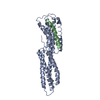 1rkcS S: Starting model for refinement C: citing same article ( |
|---|---|
| Similar structure data |


-Links
 PDBj
PDBj


- Assembly
| Deposited unit | 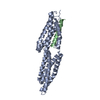
| ||||||||
|---|---|---|---|---|---|---|---|---|---|
| 1 |
| ||||||||
| 2 | 
| ||||||||
| Unit cell |
|
-Components
| #1: Protein | Mass: 28751.188 Da / Num. of mol.: 1 Source method: isolated from a genetically manipulated source Source: (gene. exp.) Homo sapiens (human) / Gene: VCL / Production host: |
|---|---|
| #2: Protein/peptide | Mass: 2616.940 Da / Num. of mol.: 1 / Fragment: unp residues 812-834 Source method: isolated from a genetically manipulated source Source: (gene. exp.) Rickettsia rickettsii (bacteria) / Strain: Iowa / Gene: RrIowa_0797 / Production host: |
| #3: Water | ChemComp-HOH /  Mass: 18.015 Da / Num. of mol.: 141 / Source method: isolated from a natural source / Formula: H2O Mass: 18.015 Da / Num. of mol.: 141 / Source method: isolated from a natural source / Formula: H2O |
-Experimental details
-Experiment
| Experiment | Method: X-RAY DIFFRACTION / Number of used crystals: 1 |
|---|
- Sample preparation
| Crystal | Density Matthews: 3.8 Å3/Da / Density % sol: 67.65 % |
|---|---|
| Crystal grow | Temperature: 298 K / Method: vapor diffusion, hanging drop / pH: 5.6 Details: 2% (v/v) tacsimate (pH 5), 100 mM sodium citrate tribasic dihydrate (pH 5.6), 16% (w/v) polyethylene glycol (3,350), 20 mM Tris-HCl (pH 9), 150 mM NaCl , VAPOR DIFFUSION, HANGING DROP, temperature 298K |
-Data collection
| Diffraction | Mean temperature: 100 K | |||||||||||||||||||||||||||||||||||||||||||||||||||||||||||||||||||||||||||||
|---|---|---|---|---|---|---|---|---|---|---|---|---|---|---|---|---|---|---|---|---|---|---|---|---|---|---|---|---|---|---|---|---|---|---|---|---|---|---|---|---|---|---|---|---|---|---|---|---|---|---|---|---|---|---|---|---|---|---|---|---|---|---|---|---|---|---|---|---|---|---|---|---|---|---|---|---|---|---|
| Diffraction source | Source: SYNCHROTRON / Site: SSRL  / Beamline: BL11-1 / Wavelength: 0.97867 Å / Beamline: BL11-1 / Wavelength: 0.97867 Å | |||||||||||||||||||||||||||||||||||||||||||||||||||||||||||||||||||||||||||||
| Detector | Type: MARMOSAIC 325 mm CCD / Detector: CCD / Date: Jul 10, 2010 | |||||||||||||||||||||||||||||||||||||||||||||||||||||||||||||||||||||||||||||
| Radiation | Monochromator: Side scattering bent cube-root I-beam single crystal; asymmetric cut 4.965 degrees Protocol: SINGLE WAVELENGTH / Monochromatic (M) / Laue (L): M / Scattering type: x-ray | |||||||||||||||||||||||||||||||||||||||||||||||||||||||||||||||||||||||||||||
| Radiation wavelength | Wavelength: 0.97867 Å / Relative weight: 1 | |||||||||||||||||||||||||||||||||||||||||||||||||||||||||||||||||||||||||||||
| Reflection | Resolution: 2.76→99.97 Å / Num. all: 12290 / Num. obs: 12290 / % possible obs: 100 % / Observed criterion σ(F): 0 / Observed criterion σ(I): 0 / Redundancy: 14.8 % / Biso Wilson estimate: 72.81 Å2 / Rmerge(I) obs: 0.059 / Net I/σ(I): 36.4 | |||||||||||||||||||||||||||||||||||||||||||||||||||||||||||||||||||||||||||||
| Reflection shell |
|
- Processing
| Software |
| |||||||||||||||||||||||||||||||||||||||||||||||||||||||||||||||||||||||||||
|---|---|---|---|---|---|---|---|---|---|---|---|---|---|---|---|---|---|---|---|---|---|---|---|---|---|---|---|---|---|---|---|---|---|---|---|---|---|---|---|---|---|---|---|---|---|---|---|---|---|---|---|---|---|---|---|---|---|---|---|---|---|---|---|---|---|---|---|---|---|---|---|---|---|---|---|---|
| Refinement | Method to determine structure: MOLECULAR REPLACEMENT Starting model: pdb entry 1rkc Resolution: 2.76→44.71 Å / Cor.coef. Fo:Fc: 0.9461 / Cor.coef. Fo:Fc free: 0.9227 / Cross valid method: THROUGHOUT / σ(F): 0
| |||||||||||||||||||||||||||||||||||||||||||||||||||||||||||||||||||||||||||
| Displacement parameters | Biso mean: 79.59 Å2
| |||||||||||||||||||||||||||||||||||||||||||||||||||||||||||||||||||||||||||
| Refine analyze | Luzzati coordinate error obs: 0.389 Å | |||||||||||||||||||||||||||||||||||||||||||||||||||||||||||||||||||||||||||
| Refinement step | Cycle: LAST / Resolution: 2.76→44.71 Å
| |||||||||||||||||||||||||||||||||||||||||||||||||||||||||||||||||||||||||||
| Refine LS restraints |
| |||||||||||||||||||||||||||||||||||||||||||||||||||||||||||||||||||||||||||
| LS refinement shell | Resolution: 2.76→3.02 Å / Total num. of bins used: 6
| |||||||||||||||||||||||||||||||||||||||||||||||||||||||||||||||||||||||||||
| Refinement TLS params. | Method: refined / Refine-ID: X-RAY DIFFRACTION
| |||||||||||||||||||||||||||||||||||||||||||||||||||||||||||||||||||||||||||
| Refinement TLS group |
|
