 Movie
Movie Controller

[English] 日本語
 Yorodumi
- PDB-4e17: Alpha-E-catenin is an autoinhibited molecule that co-activates vi... -
+ Open data

- Basic information
| Entry | Database: PDB / ID: 4.0E+17 | ||||||
|---|---|---|---|---|---|---|---|
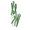
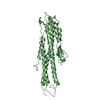
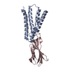
| Title | Alpha-E-catenin is an autoinhibited molecule that co-activates vinculin | ||||||
 Components Components |
| ||||||
 Keywords Keywords | CELL ADHESION / four helix bundle | ||||||
| Function / homology |  Function and homology information Function and homology informationmuscle tendon junction / Platelet degranulation / Smooth Muscle Contraction / negative regulation of integrin-mediated signaling pathway / regulation of protein localization to adherens junction / outer dense plaque of desmosome / inner dense plaque of desmosome / podosome ring / VEGFR2 mediated vascular permeability / terminal web ...muscle tendon junction / Platelet degranulation / Smooth Muscle Contraction / negative regulation of integrin-mediated signaling pathway / regulation of protein localization to adherens junction / outer dense plaque of desmosome / inner dense plaque of desmosome / podosome ring / VEGFR2 mediated vascular permeability / terminal web / Adherens junctions interactions / RHO GTPases activate IQGAPs / epithelial cell-cell adhesion / Regulation of CDH1 Function / zonula adherens / Degradation of CDH1 / Regulation of CDH1 Function / gamma-catenin binding / gap junction assembly / fascia adherens / MAP2K and MAPK activation / dystroglycan binding / alpha-catenin binding / cellular response to indole-3-methanol / muscle alpha-actinin binding / vinculin binding / Myogenesis / cell-cell contact zone / flotillin complex / apical junction assembly / costamere / regulation of establishment of endothelial barrier / catenin complex / positive regulation of extrinsic apoptotic signaling pathway in absence of ligand / negative regulation of cell motility / positive regulation of smoothened signaling pathway / axon extension / protein localization to cell surface / adherens junction assembly / axon regeneration / regulation of focal adhesion assembly / lamellipodium assembly / negative regulation of neuroblast proliferation / negative regulation of protein localization to nucleus / odontogenesis of dentin-containing tooth / establishment or maintenance of cell polarity / skeletal muscle myofibril / brush border / alpha-actinin binding / intercalated disc / negative regulation of extrinsic apoptotic signaling pathway in absence of ligand / ovarian follicle development / stress fiber / regulation of cell migration / acrosomal vesicle / Neutrophil degranulation / morphogenesis of an epithelium / adherens junction / neuromuscular junction / cell-cell adhesion / sarcolemma / response to estrogen / beta-catenin binding / male gonad development / Z disc / cell-cell junction / actin filament binding / cell junction / intracellular protein localization / regulation of cell population proliferation / cell migration / lamellipodium / actin cytoskeleton / scaffold protein binding / cytoskeleton / cell adhesion / mitochondrial inner membrane / cadherin binding / focal adhesion / ubiquitin protein ligase binding / negative regulation of apoptotic process / perinuclear region of cytoplasm / structural molecule activity / Golgi apparatus / protein homodimerization activity / protein-containing complex / identical protein binding / nucleus / plasma membrane / cytoplasm Similarity search - Function | ||||||
| Biological species |   | ||||||
| Method |  X-RAY DIFFRACTION / SYNCHROTRON / MOLECULAR REPLACEMENT / Resolution: 2.304 Å X-RAY DIFFRACTION / SYNCHROTRON / MOLECULAR REPLACEMENT / Resolution: 2.304 Å | ||||||
 Authors Authors | Choi, H.-J. / Pokutta, S. / Cadwell, G.W. / Bankston, L.A. / Liddington, R.C. / Weis, W.I. | ||||||
 Citation Citation | Journal: To be Published Title: Conformational plasticity of alpha-catenin revealed by binding interactions with vinculin Authors: Choi, H.-J. / Pokutta, S. / Bankston, L. / Liddington, R. / Weis, W.I. | ||||||
| History |
|
- Structure visualization
| Structure viewer | Molecule: MolmilJmol/JSmol |
|---|
- Downloads & links
-Download
| PDBx/mmCIF format | 4e17.cif.gz | 121.7 KB | Display | PDBx/mmCIF format |
|---|---|---|---|---|
| PDB format | pdb4e17.ent.gz | 93.6 KB | Display | PDB format |
| PDBx/mmJSON format | 4e17.json.gz | Tree view | PDBx/mmJSON format | |
| Others |  Other downloads Other downloads |
-Validation report
| Arichive directory | https://data.pdbj.org/pub/pdb/validation_reports/e1/4e17ftp://data.pdbj.org/pub/pdb/validation_reports/e1/4e17 | HTTPS FTP |
|---|
-Related structure data
| Related structure data |  4e18C  1t01S C: citing same article ( S: Starting model for refinement |
|---|---|
| Similar structure data |


-Links
 PDBj
PDBj

- Assembly
| Deposited unit | 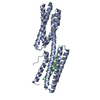
| ||||||||
|---|---|---|---|---|---|---|---|---|---|
| 1 |
| ||||||||
| Unit cell |
|
-Components
| #1: Protein | Mass: 29379.932 Da / Num. of mol.: 1 / Fragment: D1 domain (UNP residues 1-259) Source method: isolated from a genetically manipulated source Source: (gene. exp.)  |
|---|---|
| #2: Protein/peptide | Mass: 4403.830 Da / Num. of mol.: 1 / Fragment: vinculin binding domain (UNP residues 321-356) Source method: isolated from a genetically manipulated source Source: (gene. exp.) |
| #3: Water | ChemComp-HOH /  Mass: 18.015 Da / Num. of mol.: 51 / Source method: isolated from a natural source / Formula: H2O Mass: 18.015 Da / Num. of mol.: 51 / Source method: isolated from a natural source / Formula: H2O |
-Experimental details
-Experiment
| Experiment | Method: X-RAY DIFFRACTION / Number of used crystals: 1 |
|---|
- Sample preparation
| Crystal | Density Matthews: 2.69 Å3/Da / Density % sol: 54.29 % |
|---|---|
| Crystal grow | Temperature: 283 K / Method: vapor diffusion, hanging drop / pH: 6.8 Details: 20% PEG8000, 100 mM MES, pH 6.8, 3 mM DTT, VAPOR DIFFUSION, HANGING DROP, temperature 283K |
-Data collection
| Diffraction | Mean temperature: 193 K | |||||||||||||||||||||||||||||||||||||||||||||||||||||||||||||||||||||||||||||||||||||||||||||||||||||||||||||||||||||||||||||||||||||||||||||||||||
|---|---|---|---|---|---|---|---|---|---|---|---|---|---|---|---|---|---|---|---|---|---|---|---|---|---|---|---|---|---|---|---|---|---|---|---|---|---|---|---|---|---|---|---|---|---|---|---|---|---|---|---|---|---|---|---|---|---|---|---|---|---|---|---|---|---|---|---|---|---|---|---|---|---|---|---|---|---|---|---|---|---|---|---|---|---|---|---|---|---|---|---|---|---|---|---|---|---|---|---|---|---|---|---|---|---|---|---|---|---|---|---|---|---|---|---|---|---|---|---|---|---|---|---|---|---|---|---|---|---|---|---|---|---|---|---|---|---|---|---|---|---|---|---|---|---|---|---|---|
| Diffraction source | Source: SYNCHROTRON / Site: APS  / Beamline: 23-ID-D / Wavelength: 1.0332 Å / Beamline: 23-ID-D / Wavelength: 1.0332 Å | |||||||||||||||||||||||||||||||||||||||||||||||||||||||||||||||||||||||||||||||||||||||||||||||||||||||||||||||||||||||||||||||||||||||||||||||||||
| Detector | Type: MARMOSAIC 300 mm CCD / Detector: CCD / Date: Nov 7, 2011 | |||||||||||||||||||||||||||||||||||||||||||||||||||||||||||||||||||||||||||||||||||||||||||||||||||||||||||||||||||||||||||||||||||||||||||||||||||
| Radiation | Monochromator: double crystal Si(111) / Protocol: SINGLE WAVELENGTH / Monochromatic (M) / Laue (L): M / Scattering type: x-ray | |||||||||||||||||||||||||||||||||||||||||||||||||||||||||||||||||||||||||||||||||||||||||||||||||||||||||||||||||||||||||||||||||||||||||||||||||||
| Radiation wavelength | Wavelength: 1.0332 Å / Relative weight: 1 | |||||||||||||||||||||||||||||||||||||||||||||||||||||||||||||||||||||||||||||||||||||||||||||||||||||||||||||||||||||||||||||||||||||||||||||||||||
| Reflection | Resolution: 2.3→50 Å / Num. obs: 16376 / % possible obs: 97.7 % / Observed criterion σ(F): 1 / Observed criterion σ(I): 1 / Redundancy: 3.5 % / Rmerge(I) obs: 0.078 / Χ2: 1.684 / Net I/σ(I): 11.2 | |||||||||||||||||||||||||||||||||||||||||||||||||||||||||||||||||||||||||||||||||||||||||||||||||||||||||||||||||||||||||||||||||||||||||||||||||||
| Reflection shell |
|
- Processing
| Software |
| ||||||||||||||||||||||||||||||||||||||||||||||||||||||||||||||||||||||||
|---|---|---|---|---|---|---|---|---|---|---|---|---|---|---|---|---|---|---|---|---|---|---|---|---|---|---|---|---|---|---|---|---|---|---|---|---|---|---|---|---|---|---|---|---|---|---|---|---|---|---|---|---|---|---|---|---|---|---|---|---|---|---|---|---|---|---|---|---|---|---|---|---|---|
| Refinement | Method to determine structure: MOLECULAR REPLACEMENT Starting model: PDB ENTRY 1T01 Resolution: 2.304→42.133 Å / Occupancy max: 1 / Occupancy min: 0.5 / FOM work R set: 0.8356 / SU ML: 0.25 / σ(F): 1.34 / Phase error: 22.95 / Stereochemistry target values: ML
| ||||||||||||||||||||||||||||||||||||||||||||||||||||||||||||||||||||||||
| Solvent computation | Shrinkage radii: 1.11 Å / VDW probe radii: 1.3 Å / Solvent model: FLAT BULK SOLVENT MODEL / Bsol: 47.345 Å2 / ksol: 0.375 e/Å3 | ||||||||||||||||||||||||||||||||||||||||||||||||||||||||||||||||||||||||
| Displacement parameters | Biso max: 149.53 Å2 / Biso mean: 50.4 Å2 / Biso min: 18.16 Å2
| ||||||||||||||||||||||||||||||||||||||||||||||||||||||||||||||||||||||||
| Refinement step | Cycle: LAST / Resolution: 2.304→42.133 Å
| ||||||||||||||||||||||||||||||||||||||||||||||||||||||||||||||||||||||||
| Refine LS restraints |
| ||||||||||||||||||||||||||||||||||||||||||||||||||||||||||||||||||||||||
| LS refinement shell | Refine-ID: X-RAY DIFFRACTION / Total num. of bins used: 9
| ||||||||||||||||||||||||||||||||||||||||||||||||||||||||||||||||||||||||
| Refinement TLS params. | S33: 0 Å ° / Method: refined / Refine-ID: X-RAY DIFFRACTION
| ||||||||||||||||||||||||||||||||||||||||||||||||||||||||||||||||||||||||
| Refinement TLS group |
|
