 Movie
Movie Controller

[English] 日本語
 Yorodumi
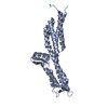
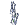
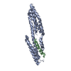
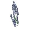
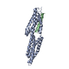
- PDB-1syq: Human vinculin head domain VH1, residues 1-258, in complex with h... -
+ Open data

- Basic information
| Entry | Database: PDB / ID: 1syq | ||||||
|---|---|---|---|---|---|---|---|
| Title | Human vinculin head domain VH1, residues 1-258, in complex with human talin's vinculin binding site 1, residues 607-636 | ||||||
 Components Components |
| ||||||
 Keywords Keywords | CELL ADHESION / cytoskeleton / vinculin / talin | ||||||
| Function / homology |  Function and homology information Function and homology informationregulation of protein localization to adherens junction / outer dense plaque of desmosome / inner dense plaque of desmosome / podosome ring / terminal web / cell-substrate junction / epithelial cell-cell adhesion / zonula adherens / LIM domain binding / fascia adherens ...regulation of protein localization to adherens junction / outer dense plaque of desmosome / inner dense plaque of desmosome / podosome ring / terminal web / cell-substrate junction / epithelial cell-cell adhesion / zonula adherens / LIM domain binding / fascia adherens / dystroglycan binding / alpha-catenin binding / vinculin binding / cortical microtubule organization / SEMA3A-Plexin repulsion signaling by inhibiting Integrin adhesion / XBP1(S) activates chaperone genes / cell-cell contact zone / integrin activation / apical junction assembly / costamere / axon extension / regulation of establishment of endothelial barrier / cell-substrate junction assembly / Regulation of CDH1 Function / cell-cell junction assembly / adherens junction assembly / cortical actin cytoskeleton organization / protein localization to cell surface / lamellipodium assembly / regulation of focal adhesion assembly / p130Cas linkage to MAPK signaling for integrins / phosphatidylserine binding / maintenance of blood-brain barrier / GRB2:SOS provides linkage to MAPK signaling for Integrins / brush border / Smooth Muscle Contraction / ruffle / Integrin signaling / phosphatidylinositol binding / Turbulent (oscillatory, disturbed) flow shear stress activates signaling by PIEZO1 and integrins in endothelial cells / negative regulation of cell migration / morphogenesis of an epithelium / integrin-mediated signaling pathway / cell-matrix adhesion / adherens junction / cell-cell adhesion / Signaling by high-kinase activity BRAF mutants / MAP2K and MAPK activation / sarcolemma / beta-catenin binding / structural constituent of cytoskeleton / platelet aggregation / integrin binding / specific granule lumen / ruffle membrane / Signaling by RAF1 mutants / cell-cell junction / Signaling by moderate kinase activity BRAF mutants / Paradoxical activation of RAF signaling by kinase inactive BRAF / Signaling downstream of RAS mutants / actin filament binding / Signaling by BRAF and RAF1 fusions / Signaling by ALK fusions and activated point mutants / Platelet degranulation / extracellular vesicle / actin cytoskeleton organization / actin binding / secretory granule lumen / High laminar flow shear stress activates signaling by PIEZO1 and PECAM1:CDH5:KDR in endothelial cells / molecular adaptor activity / ficolin-1-rich granule lumen / cytoskeleton / cell adhesion / cadherin binding / membrane raft / focal adhesion / ubiquitin protein ligase binding / Neutrophil degranulation / perinuclear region of cytoplasm / structural molecule activity / cell surface / protein-containing complex / extracellular exosome / extracellular region / plasma membrane / cytosol / cytoplasm Similarity search - Function | ||||||
| Biological species |  Homo sapiens (human) Homo sapiens (human) | ||||||
| Method |  X-RAY DIFFRACTION / SYNCHROTRON / MOLECULAR REPLACEMENT / Resolution: 2.42 Å X-RAY DIFFRACTION / SYNCHROTRON / MOLECULAR REPLACEMENT / Resolution: 2.42 Å | ||||||
 Authors Authors | Izard, T. / Vonrhein, C. | ||||||
 Citation Citation | Journal: J.Biol.Chem. / Year: 2004 Title: Structural basis for amplifying vinculin activation by talin Authors: Izard, T. / Vonrhein, C. | ||||||
| History |
|
- Structure visualization
| Structure viewer | Molecule: MolmilJmol/JSmol |
|---|
- Downloads & links
-Download
| PDBx/mmCIF format | 1syq.cif.gz | 71.6 KB | Display | PDBx/mmCIF format |
|---|---|---|---|---|
| PDB format | pdb1syq.ent.gz | 53.6 KB | Display | PDB format |
| PDBx/mmJSON format | 1syq.json.gz | Tree view | PDBx/mmJSON format | |
| Others |  Other downloads Other downloads |
-Validation report
| Arichive directory | https://data.pdbj.org/pub/pdb/validation_reports/sy/1syqftp://data.pdbj.org/pub/pdb/validation_reports/sy/1syq | HTTPS FTP |
|---|
-Related structure data
| Similar structure data |
|---|


-Links
 PDBj
PDBj


- Assembly
| Deposited unit | 
| ||||||||
|---|---|---|---|---|---|---|---|---|---|
| 1 |
| ||||||||
| 2 | x 6
| ||||||||
| 3 | x 6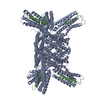
| ||||||||
| Unit cell |
|
-Components
| #1: Protein | Mass: 29766.275 Da / Num. of mol.: 1 Source method: isolated from a genetically manipulated source Source: (gene. exp.) Homo sapiens (human) / Plasmid: peT3d / Species (production host): Escherichia coli / Production host:  |
|---|---|
| #2: Protein/peptide | Mass: 2421.792 Da / Num. of mol.: 1 / Source method: obtained synthetically / References: UniProt: Q9Y490 |
| #3: Water | ChemComp-HOH /  Mass: 18.015 Da / Num. of mol.: 190 / Source method: isolated from a natural source / Formula: H2O Mass: 18.015 Da / Num. of mol.: 190 / Source method: isolated from a natural source / Formula: H2O |
-Experimental details
-Experiment
| Experiment | Method: X-RAY DIFFRACTION / Number of used crystals: 1 |
|---|
- Sample preparation
| Crystal | Density Matthews: 3.12 Å3/Da / Density % sol: 60.59 % |
|---|
-Data collection
| Diffraction | Mean temperature: 100 K |
|---|---|
| Diffraction source | Source: SYNCHROTRON / Site: APS  / Beamline: 19-ID / Wavelength: 0.97889 / Beamline: 19-ID / Wavelength: 0.97889 |
| Radiation | Protocol: SINGLE WAVELENGTH / Monochromatic (M) / Laue (L): M / Scattering type: x-ray |
| Radiation wavelength | Wavelength: 0.97889 Å / Relative weight: 1 |
| Reflection | Resolution: 2.42→50 Å / Num. obs: 15061 / % possible obs: 97.6 % / Redundancy: 18 % / Biso Wilson estimate: 34.9 Å2 / Rmerge(I) obs: 0.179 / Net I/σ(I): 8.3 |
| Reflection shell | Resolution: 2.42→2.51 Å / Rmerge(I) obs: 0.485 / Mean I/σ(I) obs: 3 / % possible all: 80.8 |
- Processing
| Software |
| ||||||||||||||||||||||||||||||||||||||||||||||||||||||||||||||||||||||||||||||||
|---|---|---|---|---|---|---|---|---|---|---|---|---|---|---|---|---|---|---|---|---|---|---|---|---|---|---|---|---|---|---|---|---|---|---|---|---|---|---|---|---|---|---|---|---|---|---|---|---|---|---|---|---|---|---|---|---|---|---|---|---|---|---|---|---|---|---|---|---|---|---|---|---|---|---|---|---|---|---|---|---|---|
| Refinement | Method to determine structure: MOLECULAR REPLACEMENT / Resolution: 2.42→32.25 Å / Rfactor Rfree error: 0.007 / Data cutoff high absF: 2709941.09 / Data cutoff low absF: 0 / Isotropic thermal model: RESTRAINED / Cross valid method: THROUGHOUT / σ(F): 0
| ||||||||||||||||||||||||||||||||||||||||||||||||||||||||||||||||||||||||||||||||
| Solvent computation | Solvent model: FLAT MODEL / Bsol: 53.5471 Å2 / ksol: 0.322653 e/Å3 | ||||||||||||||||||||||||||||||||||||||||||||||||||||||||||||||||||||||||||||||||
| Displacement parameters | Biso mean: 56.3 Å2
| ||||||||||||||||||||||||||||||||||||||||||||||||||||||||||||||||||||||||||||||||
| Refine analyze |
| ||||||||||||||||||||||||||||||||||||||||||||||||||||||||||||||||||||||||||||||||
| Refinement step | Cycle: LAST / Resolution: 2.42→32.25 Å
| ||||||||||||||||||||||||||||||||||||||||||||||||||||||||||||||||||||||||||||||||
| Refine LS restraints |
| ||||||||||||||||||||||||||||||||||||||||||||||||||||||||||||||||||||||||||||||||
| LS refinement shell | Resolution: 2.42→2.57 Å / Rfactor Rfree error: 0.024 / Total num. of bins used: 6
| ||||||||||||||||||||||||||||||||||||||||||||||||||||||||||||||||||||||||||||||||
| Xplor file |
|
