[English] 日本語
Yorodumi
- PDB-3tj5: human vinculin head domain (Vh1, residues 1-258) in complex with ... -

+
Open data


ID or keywords:

Loading...

-
Basic information

Entry
Database: PDB / ID: 3tj5
Title
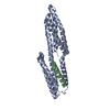
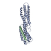
human vinculin head domain (Vh1, residues 1-258) in complex with the vinculin binding site of the surface cell antigen 4 (sca4-VBS-N; residues 412-434) from Rickettsia rickettsii
Components
  • Antigenic heat-stable 120 kDa protein
  • Vinculin
Keywordsprotein binding/toxin / cytoskeleton / epidemic typhus / sca4 / spotted fever / ALPHA-HELIX BUNDLE DOMAIN / PROTEIN-PROTEIN INTERACTIONS / CELL ADHESION / CYTOSOL / FOCAL ADHESION / protein binding-toxin complex
Function / homology
Function and homology information


regulation of protein localization to adherens junction / podosome ring / outer dense plaque of desmosome / inner dense plaque of desmosome / terminal web / cell-substrate junction / epithelial cell-cell adhesion / zonula adherens / dystroglycan binding / alpha-catenin binding ...regulation of protein localization to adherens junction / podosome ring / outer dense plaque of desmosome / inner dense plaque of desmosome / terminal web / cell-substrate junction / epithelial cell-cell adhesion / zonula adherens / dystroglycan binding / alpha-catenin binding / fascia adherens / cell-cell contact zone / adherens junction assembly / apical junction assembly / costamere / regulation of establishment of endothelial barrier / axon extension / protein localization to cell surface / lamellipodium assembly / regulation of focal adhesion assembly / maintenance of blood-brain barrier / brush border / Smooth Muscle Contraction / negative regulation of cell migration / cell-matrix adhesion / cell projection / morphogenesis of an epithelium / adherens junction / Signaling by high-kinase activity BRAF mutants / MAP2K and MAPK activation / sarcolemma / beta-catenin binding / platelet aggregation / specific granule lumen / Signaling by RAF1 mutants / Signaling by moderate kinase activity BRAF mutants / Paradoxical activation of RAF signaling by kinase inactive BRAF / Signaling downstream of RAS mutants / Signaling by BRAF and RAF1 fusions / cell-cell junction / extracellular vesicle / Signaling by ALK fusions and activated point mutants / Platelet degranulation / actin binding / secretory granule lumen / ficolin-1-rich granule lumen / molecular adaptor activity / cytoskeleton / cell adhesion / cadherin binding / membrane raft / focal adhesion / ubiquitin protein ligase binding / Neutrophil degranulation / structural molecule activity / protein-containing complex / extracellular exosome / extracellular region / plasma membrane / cytoplasm / cytosol
Similarity search - Function
Rickettsia surface antigen, 120kDa / 120 KDa Rickettsia surface antigen / Vinculin repeated domain signature. / Vinculin / Alpha-catenin/vinculin-like / Vinculin, conserved site / Vinculin family talin-binding region signature. / Vinculin/alpha-catenin / Vinculin family / Alpha-catenin/vinculin-like superfamily ...Rickettsia surface antigen, 120kDa / 120 KDa Rickettsia surface antigen / Vinculin repeated domain signature. / Vinculin / Alpha-catenin/vinculin-like / Vinculin, conserved site / Vinculin family talin-binding region signature. / Vinculin/alpha-catenin / Vinculin family / Alpha-catenin/vinculin-like superfamily / Four Helix Bundle (Hemerythrin (Met), subunit A) / Up-down Bundle / Mainly Alpha
Similarity search - Domain/homology
Antigenic heat-stable 120 kDa protein / Vinculin
Similarity search - Component
Biological speciesHomo sapiens (human)
Rickettsia rickettsii str. Iowa (bacteria)
MethodX-RAY DIFFRACTION / SYNCHROTRON / MOLECULAR REPLACEMENT / Resolution: 1.99 Å
AuthorsPark, H. / Lee, J.H. / Gouin, E. / Cossart, P. / Izard, T.
CitationJournal: J.Biol.Chem. / Year: 2011
Title: The rickettsia surface cell antigen 4 applies mimicry to bind to and activate vinculin.
Authors: Park, H. / Lee, J.H. / Gouin, E. / Cossart, P. / Izard, T.
History
DepositionAug 23, 2011Deposition site: RCSB / Processing site: RCSB
Revision 1.0Sep 7, 2011Provider: repository / Type: Initial release
Revision 1.1Nov 2, 2011Group: Database references
Revision 1.2May 1, 2013Group: Other
Revision 1.3Nov 8, 2017Group: Refinement description / Category: software / Item: _software.name
Revision 1.4Sep 13, 2023Group: Data collection / Database references ...Data collection / Database references / Derived calculations / Refinement description
Category: chem_comp_atom / chem_comp_bond ...chem_comp_atom / chem_comp_bond / database_2 / pdbx_initial_refinement_model / software / struct_ref_seq_dif / struct_site
Item: _database_2.pdbx_DOI / _database_2.pdbx_database_accession ..._database_2.pdbx_DOI / _database_2.pdbx_database_accession / _software.name / _struct_ref_seq_dif.details / _struct_site.pdbx_auth_asym_id / _struct_site.pdbx_auth_comp_id / _struct_site.pdbx_auth_seq_id

-
Structure visualization

Structure viewerMolecule:
MolmilJmol/JSmol

Downloads & links

-
Assembly

Deposited unit
A: Vinculin
B: Antigenic heat-stable 120 kDa protein
hetero molecules


Theoretical massNumber of molelcules
Total (without water)31,4693
Polymers31,3772
Non-polymers921
Water5,693316
1


  • Idetical with deposited unit
  • defined by author&software
TypeNameSymmetry operationNumber
identity operation1_555x,y,z1
Buried area2410 Å2
ΔGint-28 kcal/mol
Surface area15080 Å2
MethodPISA
Unit cell
Length a, b, c (Å)54.404, 76.343, 114.549
Angle α, β, γ (deg.)90.00, 90.00, 90.00
Int Tables number19
Space group name H-MP212121

-
Components

#1: Protein Vinculin / Metavinculin


Mass: 28565.021 Da / Num. of mol.: 1 / Fragment: unp residues 1-255
Source method: isolated from a genetically manipulated source
Source: (gene. exp.) Homo sapiens (human) / Gene: VCL / Production host: Escherichia coli (E. coli) / References: UniProt: P18206
#2: Protein/peptide Antigenic heat-stable 120 kDa protein


Mass: 2812.165 Da / Num. of mol.: 1 / Fragment: unp residues 412-434
Source method: isolated from a genetically manipulated source
Source: (gene. exp.) Rickettsia rickettsii str. Iowa (bacteria)
Strain: Iowa / Gene: RrIowa_0797, sca4 / Production host: Escherichia coli (E. coli) / References: UniProt: B0BXR4
#3: Chemical ChemComp-GOL / GLYCEROL / GLYCERIN / PROPANE-1,2,3-TRIOL


Mass: 92.094 Da / Num. of mol.: 1 / Source method: obtained synthetically / Formula: C3H8O3
#4: Water ChemComp-HOH / water


Mass: 18.015 Da / Num. of mol.: 316 / Source method: isolated from a natural source / Formula: H2O

-
Experimental details

-
Experiment

ExperimentMethod: X-RAY DIFFRACTION / Number of used crystals: 1

-
Sample preparation

CrystalDensity Matthews: 3.79 Å3/Da / Density % sol: 67.55 %
Crystal growTemperature: 298 K / pH: 6.5
Details: 0.1 M MES monohydrate pH 6.5, 14% PEG 20,000, 20 mM Tris-HCl pH 8, 150 mM NaCl, VAPOR DIFFUSION, HANGING DROP, temperature 298K

-
Data collection

Diffraction
IDMean temperature (K)Crystal-ID
11001
21
Diffraction source
SourceSiteBeamlineIDWavelength
SYNCHROTRONSSRL BL11-111
2
Detector
TypeIDDetectorDate
MARMOSAIC 325 mm CCD1CCDJul 10, 2010
2
Radiation
IDMonochromatorProtocolMonochromatic (M) / Laue (L)Scattering typeWavelength-ID
1SIDE SCATTERING BENT CUBE-ROOT I -BEAM SINGLE CRYSTALSINGLE WAVELENGTHMx-ray1
2ASYMMETRIC CUT 4.965 DEGREESx-ray1
Radiation wavelengthWavelength: 1 Å / Relative weight: 1
ReflectionResolution: 1.99→114.55 Å / Num. obs: 33356 / % possible obs: 99.9 % / Observed criterion σ(I): 0 / Redundancy: 5.9 % / Biso Wilson estimate: 39.37 Å2 / Rmerge(I) obs: 0.034 / Net I/σ(I): 26.3
Reflection shellResolution: 1.99→2.1 Å / Redundancy: 5.3 % / Rmerge(I) obs: 0.499 / % possible all: 100

-
Processing

Software
NameVersionClassification
Blu-IceICEdata collection
MOLREPphasing
BUSTER2.9.2refinement
autoPROCdata scaling
RefinementMethod to determine structure: MOLECULAR REPLACEMENT
Starting model: pdb entry 1rkc
Resolution: 1.99→23.39 Å / Cor.coef. Fo:Fc: 0.942 / Cor.coef. Fo:Fc free: 0.933 / Cross valid method: THROUGHOUT / σ(F): 0
RfactorNum. reflection% reflectionSelection details
Rfree0.218 1681 5.05 %RANDOM
Rwork0.194 ---
obs0.195 33271 --
all-33356 --
Displacement parametersBiso mean: 52.66 Å2
Baniso -1Baniso -2Baniso -3
1--10.8495 Å20 Å20 Å2
2---2.4525 Å20 Å2
3---13.302 Å2
Refine analyzeLuzzati coordinate error obs: 0.27 Å
Refinement stepCycle: LAST / Resolution: 1.99→23.39 Å
ProteinNucleic acidLigandSolventTotal
Num. atoms2176 0 6 316 2498
Refine LS restraints
Refine-IDTypeDev idealNumberRestraint functionWeight
X-RAY DIFFRACTIONt_bond_d0.012304HARMONIC2
X-RAY DIFFRACTIONt_angle_deg0.993108HARMONIC2
X-RAY DIFFRACTIONt_dihedral_angle_d
X-RAY DIFFRACTIONt_incorr_chiral_ct
X-RAY DIFFRACTIONt_pseud_angle
X-RAY DIFFRACTIONt_trig_c_planes
X-RAY DIFFRACTIONt_gen_planes
X-RAY DIFFRACTIONt_it
X-RAY DIFFRACTIONt_nbd
X-RAY DIFFRACTIONt_omega_torsion2.33
X-RAY DIFFRACTIONt_other_torsion2.9
X-RAY DIFFRACTIONt_improper_torsion
X-RAY DIFFRACTIONt_chiral_improper_torsion
X-RAY DIFFRACTIONt_sum_occupancies
X-RAY DIFFRACTIONt_utility_distance
X-RAY DIFFRACTIONt_utility_angle
X-RAY DIFFRACTIONt_utility_torsion
X-RAY DIFFRACTIONt_ideal_dist_contact
LS refinement shellResolution: 1.99→2.05 Å / Total num. of bins used: 17
RfactorNum. reflection% reflection
Rfree0.2601 129 4.74 %
Rwork0.2349 2590 -
all0.2361 2719 -
Refinement TLS params.

Method: refined / Refine-ID: X-RAY DIFFRACTION

IDL112)L122)L132)L222)L232)L332)S11 (Å °)S12 (Å °)S13 (Å °)S21 (Å °)S22 (Å °)S23 (Å °)S31 (Å °)S32 (Å °)S33 (Å °)T112)T122)T132)T222)T232)T332)Origin x (Å)Origin y (Å)Origin z (Å)
10.2470.6613-0.70791.3834-2.45393.8457-0.14030.0558-0.0488-0.2660.1263-0.0730.4492-0.17120.014-0.012300.0504-0.1003-0.0088-0.1004-25.0953-6.477918.7588
23.2336-0.9468-0.27891.5875-2.08914.6145-0.00380.17850.30930.024-0.0081-0.229-0.39090.06350.01190.0311-0.052-0.0424-0.03380.0108-0.0302-21.8824-7.214840.4398
Refinement TLS group
IDRefine-IDRefine TLS-IDSelection detailsAuth asym-IDAuth seq-ID
1X-RAY DIFFRACTION1{ A|2 - A|255 }A2 - 255
2X-RAY DIFFRACTION2{ B|408 - B|434 }B408 - 434

+
About Yorodumi

-
News

-
Feb 9, 2022. New format data for meta-information of EMDB entries

New format data for meta-information of EMDB entries

  • Version 3 of the EMDB header file is now the official format.
  • The previous official version 1.9 will be removed from the archive.

Related info.:EMDB header

External links:wwPDB to switch to version 3 of the EMDB data model

-
Aug 12, 2020. Covid-19 info

Covid-19 info

URL: https://pdbjlvh1.pdbj.org/emnavi/covid19.php

New page: Covid-19 featured information page in EM Navigator.

Related info.:Covid-19 info / Mar 5, 2020. Novel coronavirus structure data

+
Mar 5, 2020. Novel coronavirus structure data

Novel coronavirus structure data

Related info.:Yorodumi Speices / Aug 12, 2020. Covid-19 info

External links:COVID-19 featured content - PDBj / Molecule of the Month (242):Coronavirus Proteases

+
Jan 31, 2019. EMDB accession codes are about to change! (news from PDBe EMDB page)

EMDB accession codes are about to change! (news from PDBe EMDB page)

  • The allocation of 4 digits for EMDB accession codes will soon come to an end. Whilst these codes will remain in use, new EMDB accession codes will include an additional digit and will expand incrementally as the available range of codes is exhausted. The current 4-digit format prefixed with “EMD-” (i.e. EMD-XXXX) will advance to a 5-digit format (i.e. EMD-XXXXX), and so on. It is currently estimated that the 4-digit codes will be depleted around Spring 2019, at which point the 5-digit format will come into force.
  • The EM Navigator/Yorodumi systems omit the EMD- prefix.

Related info.:Q: What is EMD? / ID/Accession-code notation in Yorodumi/EM Navigator

External links:EMDB Accession Codes are Changing Soon! / Contact to PDBj

+
Jul 12, 2017. Major update of PDB

Major update of PDB

  • wwPDB released updated PDB data conforming to the new PDBx/mmCIF dictionary.
  • This is a major update changing the version number from 4 to 5, and with Remediation, in which all the entries are updated.
  • In this update, many items about electron microscopy experimental information are reorganized (e.g. em_software).
  • Now, EM Navigator and Yorodumi are based on the updated data.

External links:wwPDB Remediation / Enriched Model Files Conforming to OneDep Data Standards Now Available in the PDB FTP Archive

-
Yorodumi

Thousand views of thousand structures

  • Yorodumi is a browser for structure data from EMDB, PDB, SASBDB, etc.
  • This page is also the successor to EM Navigator detail page, and also detail information page/front-end page for Omokage search.
  • The word "yorodu" (or yorozu) is an old Japanese word meaning "ten thousand". "mi" (miru) is to see.

Related info.:EMDB / PDB / SASBDB / Comparison of 3 databanks / Yorodumi Search / Aug 31, 2016. New EM Navigator & Yorodumi / Yorodumi Papers / Jmol/JSmol / Function and homology information / Changes in new EM Navigator and Yorodumi

Read more