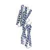
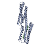
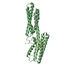
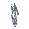
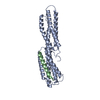
Entry Database : PDB / ID : 6fq4Title Structure of Chlamydial virulence factor TarP and vinculin head domain Keywords / / / / Function / homology Function Domain/homology Component
/ / / / / / / / / / / / / / / / / / / / / / / / / / / / / / / / / / / / / / / / / / / / / / / / / / / / / / / / / / / / / / / / / / / / / / / / / / / / / / / / / / / Biological species Gallus gallus (chicken)Chlamydia caviae (bacteria)Method / / / Resolution : 2.89 Å Authors Whitewood, A.J. / Singh, A.K. / Brown, D.G. / Goult, B.T. Funding support Organization Grant number Country Biotechnology and Biological Sciences Research Council BB/N007336/1 Human Frontier Science Program (HFSP) RGP00001/2016
Journal : FEBS Lett. / Year : 2018Title : Chlamydial virulence factor TarP mimics talin to disrupt the talin-vinculin complex.Authors : Whitewood, A.J. / Singh, A.K. / Brown, D.G. / Goult, B.T. History Deposition Feb 13, 2018 Deposition site / Processing site Revision 1.0 May 9, 2018 Provider / Type Revision 1.1 Jun 13, 2018 Group / Database references / Category / citation_authorItem _citation.journal_volume / _citation.page_first ... _citation.journal_volume / _citation.page_first / _citation.page_last / _citation_author.name Revision 1.2 Jan 17, 2024 Group / Database references / Refinement descriptionCategory chem_comp_atom / chem_comp_bond ... chem_comp_atom / chem_comp_bond / database_2 / pdbx_initial_refinement_model Item / _database_2.pdbx_database_accession
Show all Show less
 Movie
Movie Controller

 Yorodumi
 Open data
 Basic information
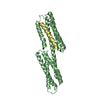
 Components
 Keywords
 Function and homology information
 Chlamydia caviae (bacteria)
 X-RAY DIFFRACTION /
  Authors
 United Kingdom, 2items
  Citation
 Structure visualization
 Downloads & links
 Other downloads


 PDBj
PDBj
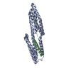
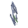
 Assembly
 Mass: 18.015 Da / Num. of mol.: 3 / Source method: isolated from a natural source / Formula: H2O
 Sample preparation
 Processing