 Movie
Movie Controller

[English] 日本語
 Yorodumi
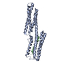
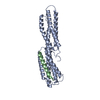
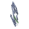
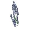
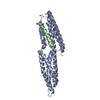
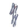
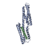
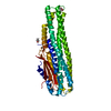
- PDB-1zvz: Vinculin Head (0-258) in Complex with the Talin Rod Residue 820-844 -
+ Open data

- Basic information
| Entry | Database: PDB / ID: 1zvz | ||||||
|---|---|---|---|---|---|---|---|
| Title | Vinculin Head (0-258) in Complex with the Talin Rod Residue 820-844 | ||||||
 Components Components |
| ||||||
 Keywords Keywords | PROTEIN BINDING / talin / vinculin / complex | ||||||
| Function / homology |  Function and homology information Function and homology informationmuscle tendon junction / Platelet degranulation / Smooth Muscle Contraction / regulation of protein localization to adherens junction / outer dense plaque of desmosome / inner dense plaque of desmosome / podosome ring / terminal web / epithelial cell-cell adhesion / zonula adherens ...muscle tendon junction / Platelet degranulation / Smooth Muscle Contraction / regulation of protein localization to adherens junction / outer dense plaque of desmosome / inner dense plaque of desmosome / podosome ring / terminal web / epithelial cell-cell adhesion / zonula adherens / Regulation of CDH1 Function / fascia adherens / MAP2K and MAPK activation / dystroglycan binding / alpha-catenin binding / muscle alpha-actinin binding / vinculin binding / cell-cell contact zone / apical junction assembly / costamere / regulation of establishment of endothelial barrier / axon extension / protein localization to cell surface / adherens junction assembly / regulation of focal adhesion assembly / lamellipodium assembly / skeletal muscle myofibril / brush border / alpha-actinin binding / ruffle / stress fiber / regulation of cell migration / Neutrophil degranulation / morphogenesis of an epithelium / adherens junction / neuromuscular junction / cell-cell adhesion / sarcolemma / structural constituent of cytoskeleton / beta-catenin binding / integrin binding / ruffle membrane / Z disc / cell-cell junction / actin filament binding / actin cytoskeleton / actin cytoskeleton organization / scaffold protein binding / cytoskeleton / cell adhesion / mitochondrial inner membrane / cadherin binding / focal adhesion / ubiquitin protein ligase binding / perinuclear region of cytoplasm / structural molecule activity / cell surface / protein homodimerization activity / protein-containing complex / plasma membrane / cytoplasm / cytosol Similarity search - Function | ||||||
| Biological species |  | ||||||
| Method |  X-RAY DIFFRACTION / SYNCHROTRON / MOLECULAR REPLACEMENT / Resolution: 1.8 Å X-RAY DIFFRACTION / SYNCHROTRON / MOLECULAR REPLACEMENT / Resolution: 1.8 Å | ||||||
 Authors Authors | Gingras, A.R. / Ziegler, W.H. / Barsukov, I.L. / Roberts, G.C. / Critchley, D.R. / Emsley, J. | ||||||
 Citation Citation | Journal: J.Biol.Chem. / Year: 2005 Title: Mapping and consensus sequence identification for multiple vinculin binding sites within the talin rod Authors: Gingras, A.R. / Ziegler, W.H. / Frank, R. / Barsukov, I.L. / Roberts, G.C. / Critchley, D.R. / Emsley, J. | ||||||
| History |
|
- Structure visualization
| Structure viewer | Molecule: MolmilJmol/JSmol |
|---|
- Downloads & links
-Download
| PDBx/mmCIF format | 1zvz.cif.gz | 71.5 KB | Display | PDBx/mmCIF format |
|---|---|---|---|---|
| PDB format | pdb1zvz.ent.gz | 51.6 KB | Display | PDB format |
| PDBx/mmJSON format | 1zvz.json.gz | Tree view | PDBx/mmJSON format | |
| Others |  Other downloads Other downloads |
-Validation report
| Arichive directory | https://data.pdbj.org/pub/pdb/validation_reports/zv/1zvzftp://data.pdbj.org/pub/pdb/validation_reports/zv/1zvz | HTTPS FTP |
|---|
-Related structure data
| Related structure data |  1zw2C  1zw3C 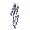 1xwjS S: Starting model for refinement C: citing same article ( |
|---|---|
| Similar structure data |


-Links
 PDBj
PDBj
- Assembly
| Deposited unit | 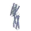
| ||||||||
|---|---|---|---|---|---|---|---|---|---|
| 1 |
| ||||||||
| Unit cell |
|
-Components
| #1: Protein | Mass: 31195.908 Da / Num. of mol.: 1 / Fragment: Residues 0-258 Source method: isolated from a genetically manipulated source Source: (gene. exp.)  |
|---|---|
| #2: Protein/peptide | Mass: 2645.021 Da / Num. of mol.: 1 / Fragment: Residues 820-844 / Source method: obtained synthetically Details: THE PEPTIDE WAS CHEMICALLY SYNTHESIZED. THE SEQUENCE OF THE PEPTIDE IS NATURALLY FOUND IN GALLUS GALLUS(CHICKEN). References: UniProt: P54939 |
| #3: Water | ChemComp-HOH /  Mass: 18.015 Da / Num. of mol.: 251 / Source method: isolated from a natural source / Formula: H2O Mass: 18.015 Da / Num. of mol.: 251 / Source method: isolated from a natural source / Formula: H2O |
-Experimental details
-Experiment
| Experiment | Method: X-RAY DIFFRACTION / Number of used crystals: 1 |
|---|
- Sample preparation
| Crystal | Density Matthews: 2.1 Å3/Da / Density % sol: 42 % |
|---|---|
| Crystal grow | Temperature: 293 K / Method: vapor diffusion, sitting drop / Details: VAPOR DIFFUSION, SITTING DROP, temperature 293K |
-Data collection
| Diffraction | Mean temperature: 100 K |
|---|---|
| Diffraction source | Source: SYNCHROTRON / Site: ESRF  / Beamline: ID14-2 / Wavelength: 0.934 / Wavelength: 0.934 Å / Beamline: ID14-2 / Wavelength: 0.934 / Wavelength: 0.934 Å |
| Detector | Type: ADSC QUANTUM 4 / Detector: CCD / Date: Mar 13, 2005 / Details: TOROIDAL ZERODUR MIRROR |
| Radiation | Monochromator: Diamond (111), Ge(220) / Protocol: SINGLE WAVELENGTH / Monochromatic (M) / Laue (L): M / Scattering type: x-ray |
| Radiation wavelength | Wavelength: 0.934 Å / Relative weight: 1 |
| Reflection | Resolution: 1.8→30 Å / Num. all: 27873 / % possible obs: 97.7 % / Rmerge(I) obs: 0.078 / Net I/σ(I): 16.1 |
| Reflection shell | Resolution: 1.8→1.86 Å / Rmerge(I) obs: 0.346 / Mean I/σ(I) obs: 2.09 / % possible all: 95.4 |
- Processing
| Software |
| |||||||||||||||||||||||||||||||||||||||||||||||||||||||||||||||||||||||||||||||||||||||||||||||||||||||||||||||||||||||||||||
|---|---|---|---|---|---|---|---|---|---|---|---|---|---|---|---|---|---|---|---|---|---|---|---|---|---|---|---|---|---|---|---|---|---|---|---|---|---|---|---|---|---|---|---|---|---|---|---|---|---|---|---|---|---|---|---|---|---|---|---|---|---|---|---|---|---|---|---|---|---|---|---|---|---|---|---|---|---|---|---|---|---|---|---|---|---|---|---|---|---|---|---|---|---|---|---|---|---|---|---|---|---|---|---|---|---|---|---|---|---|---|---|---|---|---|---|---|---|---|---|---|---|---|---|---|---|---|
| Refinement | Method to determine structure: MOLECULAR REPLACEMENT Starting model: PDB ENTRY 1XWJ Resolution: 1.8→26.29 Å / Cor.coef. Fo:Fc: 0.926 / Cor.coef. Fo:Fc free: 0.895 / SU B: 6.398 / SU ML: 0.115 / Cross valid method: THROUGHOUT / σ(F): 0 / ESU R: 0.169 / ESU R Free: 0.155 / Stereochemistry target values: MAXIMUM LIKELIHOOD / Details: HYDROGENS HAVE BEEN ADDED IN THE RIDING POSITIONS
| |||||||||||||||||||||||||||||||||||||||||||||||||||||||||||||||||||||||||||||||||||||||||||||||||||||||||||||||||||||||||||||
| Solvent computation | Ion probe radii: 0.8 Å / Shrinkage radii: 0.8 Å / VDW probe radii: 1.2 Å / Solvent model: MASK | |||||||||||||||||||||||||||||||||||||||||||||||||||||||||||||||||||||||||||||||||||||||||||||||||||||||||||||||||||||||||||||
| Displacement parameters | Biso mean: 29.204 Å2
| |||||||||||||||||||||||||||||||||||||||||||||||||||||||||||||||||||||||||||||||||||||||||||||||||||||||||||||||||||||||||||||
| Refinement step | Cycle: LAST / Resolution: 1.8→26.29 Å
| |||||||||||||||||||||||||||||||||||||||||||||||||||||||||||||||||||||||||||||||||||||||||||||||||||||||||||||||||||||||||||||
| Refine LS restraints |
| |||||||||||||||||||||||||||||||||||||||||||||||||||||||||||||||||||||||||||||||||||||||||||||||||||||||||||||||||||||||||||||
| LS refinement shell | Resolution: 1.8→1.847 Å / Total num. of bins used: 20
|
