 Movie
Movie Controller


+ Open data

- Basic information
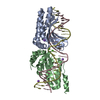
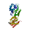
| Entry | Database: PDB / ID: 3dwo | ||||||
|---|---|---|---|---|---|---|---|
| Title | Crystal structure of a Pseudomonas aeruginosa FadL homologue | ||||||
 Components Components | Probable outer membrane protein | ||||||
 Keywords Keywords | MEMBRANE PROTEIN / beta barrel / outer membrane protein | ||||||
| Function / homology | Outer membrane protein transport protein (OMPP1/FadL/TodX) / long-chain fatty acid transporting porin activity / Outer membrane protein transport protein (OMPP1/FadL/TodX) / Outer membrane protein transport protein (OMPP1/FadL/TodX) / Porin / cell outer membrane / Beta Barrel / Mainly Beta / Probable outer membrane protein Function and homology information Function and homology information | ||||||
| Biological species |   Pseudomonas aeruginosa (bacteria) Pseudomonas aeruginosa (bacteria) | ||||||
| Method |  X-RAY DIFFRACTION / SYNCHROTRON / MIR / Resolution: 2.2 Å X-RAY DIFFRACTION / SYNCHROTRON / MIR / Resolution: 2.2 Å | ||||||
 Authors Authors | Hearn, E.M. / Patel, D.R. / Lepore, B.W. / Indic, M. / van den Berg, B. | ||||||
 Citation Citation | Journal: Nature / Year: 2009 Title: Transmembrane passage of hydrophobic compounds through a protein channel wall. Authors: Hearn, E.M. / Patel, D.R. / Lepore, B.W. / Indic, M. / van den Berg, B. #1: Journal: Science / Year: 2004Title: Crystal structure of the long-chain fatty acid transporter FadL Authors: van den Berg, B. / Black, P.N. / Clemons Jr., W.M. / Rapoport, T.A. | ||||||
| History |
|
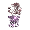
- Structure visualization
| Structure viewer | Molecule: MolmilJmol/JSmol |
|---|
- Downloads & links
-Download
| PDBx/mmCIF format | 3dwo.cif.gz | 105.7 KB | Display | PDBx/mmCIF format |
|---|---|---|---|---|
| PDB format | pdb3dwo.ent.gz | 80.7 KB | Display | PDB format |
| PDBx/mmJSON format | 3dwo.json.gz | Tree view | PDBx/mmJSON format | |
| Others |  Other downloads Other downloads |
-Validation report
| Arichive directory | https://data.pdbj.org/pub/pdb/validation_reports/dw/3dwoftp://data.pdbj.org/pub/pdb/validation_reports/dw/3dwo | HTTPS FTP |
|---|
-Related structure data
| Related structure data |  2r4lC  2r4nC  2r4oC  2r4pC  2r88C  3dwnC C: citing same article ( |
|---|---|
| Similar structure data |


-Links
 PDBj
PDBj- Assembly
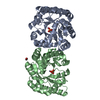
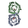
| Deposited unit | 
| ||||||||
|---|---|---|---|---|---|---|---|---|---|
| 1 |
| ||||||||
| Unit cell |
|
-Components
| #1: Protein | Mass: 48913.176 Da / Num. of mol.: 1 / Fragment: UNP residues 22-463 Source method: isolated from a genetically manipulated source Source: (gene. exp.) Pseudomonas aeruginosa (bacteria) / Strain: PAO1 / Gene: PA4589 / Plasmid: pBAD22 / Production host: | ||
|---|---|---|---|
| #2: Chemical | ChemComp-SO4 /   Mass: 96.063 Da / Num. of mol.: 1 / Source method: obtained synthetically / Formula: SO4 Mass: 96.063 Da / Num. of mol.: 1 / Source method: obtained synthetically / Formula: SO4 | ||
| #3: Chemical | ChemComp-C8E / (   Mass: 306.438 Da / Num. of mol.: 11 / Source method: obtained synthetically / Formula: C16H34O5 / Comment: C8E, detergent*YM Mass: 306.438 Da / Num. of mol.: 11 / Source method: obtained synthetically / Formula: C16H34O5 / Comment: C8E, detergent*YM#4: Water | ChemComp-HOH / |  Mass: 18.015 Da / Num. of mol.: 214 / Source method: isolated from a natural source / Formula: H2O Mass: 18.015 Da / Num. of mol.: 214 / Source method: isolated from a natural source / Formula: H2O |
-Experimental details
-Experiment
| Experiment | Method: X-RAY DIFFRACTION / Number of used crystals: 1 |
|---|
- Sample preparation
| Crystal | Density Matthews: 2.77 Å3/Da / Density % sol: 55.59 % |
|---|---|
| Crystal grow | Temperature: 295 K / Method: vapor diffusion, hanging drop / pH: 6.5 Details: 28-32% PEG 400, 0.1M lithium sulfate, 0.1M MES, pH 6.5, VAPOR DIFFUSION, HANGING DROP, temperature 295K |
-Data collection
| Diffraction | Mean temperature: 100 K | ||||||||||||
|---|---|---|---|---|---|---|---|---|---|---|---|---|---|
| Diffraction source | Source: SYNCHROTRON / Site: NSLS  / Beamline: X29A / Wavelength: 0.9790, 1.0375, 1.1397 / Beamline: X29A / Wavelength: 0.9790, 1.0375, 1.1397 | ||||||||||||
| Detector | Type: ADSC QUANTUM 315 / Detector: CCD / Date: Nov 29, 2007 | ||||||||||||
| Radiation | Monochromator: Si(111) / Protocol: SINGLE WAVELENGTH / Monochromatic (M) / Laue (L): M / Scattering type: x-ray | ||||||||||||
| Radiation wavelength |
| ||||||||||||
| Reflection | Resolution: 2.1→50 Å / Num. all: 31016 / Num. obs: 28897 / % possible obs: 89.7 % / Observed criterion σ(F): 0 / Observed criterion σ(I): 2.4 / Redundancy: 5.6 % / Rmerge(I) obs: 0.073 / Net I/av σ(I): 20.4 / Net I/σ(I): 10 | ||||||||||||
| Reflection shell | Resolution: 2.1→2.18 Å / Redundancy: 3.2 % / Rmerge(I) obs: 0.287 / Mean I/σ(I) obs: 2.4 / Num. unique all: 1218 / Rsym value: 0.287 / % possible all: 38.6 |
- Processing
| Software |
| ||||||||||||||||||||||||||||
|---|---|---|---|---|---|---|---|---|---|---|---|---|---|---|---|---|---|---|---|---|---|---|---|---|---|---|---|---|---|
| Refinement | Method to determine structure: MIR / Resolution: 2.2→20 Å / Occupancy max: 1 / Occupancy min: 1 / Cross valid method: THROUGHOUT / σ(F): 0 / σ(I): 0 / Stereochemistry target values: Engh & Huber
| ||||||||||||||||||||||||||||
| Solvent computation | Bsol: 65.517 Å2 | ||||||||||||||||||||||||||||
| Displacement parameters | Biso max: 79 Å2 / Biso mean: 33.501 Å2 / Biso min: 13.37 Å2
| ||||||||||||||||||||||||||||
| Refinement step | Cycle: LAST / Resolution: 2.2→20 Å
| ||||||||||||||||||||||||||||
| Refine LS restraints |
| ||||||||||||||||||||||||||||
| Xplor file |
|
