- PDB-5lld: Flavodiiron core of Escherichia coli flavorubredoxin in the reduc... -
+
Open data
ID or keywords:
Loading...
-
Basic information
Entry
Database: PDB / ID: 5lld
Title
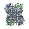
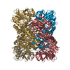
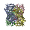
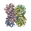
Flavodiiron core of Escherichia coli flavorubredoxin in the reduced form.
Components
Anaerobic nitric oxide reductase flavorubredoxin
Keywords
OXIDOREDUCTASE / Flavorubredoxin / flavodiiron protein / diiron center / nitric oxide reductase
Function / homology
Function and homology information
nitric oxide reductase activity / oxidoreductase activity, acting on other nitrogenous compounds as donors / nitric oxide catabolic process / response to nitric oxide / FMN binding / electron transfer activity / oxidoreductase activity / iron ion binding / protein-containing complex / identical protein binding / cytoplasm Similarity search - Function
Mass: 18.015 Da / Num. of mol.: 128 / Source method: isolated from a natural source / Formula: H2O
-
Experimental details
-
Experiment
Experiment
Method: X-RAY DIFFRACTION / Number of used crystals: 1
-
Sample preparation
Crystal
Density Matthews: 2.95 Å3/Da / Density % sol: 58.33 %
Crystal grow
Temperature: 304 K / Method: vapor diffusion, hanging drop / pH: 6.5 Details: 1.0UL OF PROTEIN AT15 MG/ML WITH 0.8UL OF CRYSTALLIZATION SOLUTION (0.2M NA-CACODYLATE PH 6.5, 0.2 M MGACETATE, 20% PEG8000) and 0.2UL OF 0.1M HEXAMINE COBALT (III).
Resolution: 2.651→45.292 Å / SU ML: 0.36 / Cross valid method: FREE R-VALUE / σ(F): 0.35 / Phase error: 13.9 / Stereochemistry target values: ML Details: The final model was refined versus the full data, resulting in R value of 0.19. The rubredoxin domain in C-terminal was not visible since it is disordered.
Rfactor
Num. reflection
% reflection
Selection details
Rfree
0.246
17132
10 %
random
Rwork
0.185
-
-
-
obs
0.1899
1713
87.72 %
-
Solvent computation
Shrinkage radii: 0.9 Å / VDW probe radii: 1.11 Å / Solvent model: FLAT BULK SOLVENT MODEL / Bsol: 0.438 Å2 / ksol: 0.297 e/Å3
Displacement parameters
Baniso -1
Baniso -2
Baniso -3
1-
-9.168 Å2
0 Å2
-0 Å2
2-
-
-9.168 Å2
-0 Å2
3-
-
-
-2.4335 Å2
Refinement step
Cycle: LAST / Resolution: 2.651→45.292 Å
Protein
Nucleic acid
Ligand
Solvent
Total
Num. atoms
3200
0
35
128
3363
Refine LS restraints
Refine-ID
Type
Dev ideal
Number
X-RAY DIFFRACTION
f_bond_d
0.015
3318
X-RAY DIFFRACTION
f_angle_d
0.566
4494
X-RAY DIFFRACTION
f_dihedral_angle_d
10.846
1198
X-RAY DIFFRACTION
f_chiral_restr
0.044
489
X-RAY DIFFRACTION
f_plane_restr
0.002
581
LS refinement shell
Resolution (Å)
Rfactor Rfree
Num. reflection Rfree
Rfactor Rwork
Num. reflection Rwork
Refine-ID
% reflection obs (%)
2.6512-2.6813
0.241
495
0.241
495
X-RAY DIFFRACTION
77
2.6813-2.7128
0.2263
511
0.2263
511
X-RAY DIFFRACTION
81
2.7128-2.7459
0.242
532
0.242
532
X-RAY DIFFRACTION
84
2.7459-2.7807
0.2198
531
0.2198
531
X-RAY DIFFRACTION
85
2.7807-2.8173
0.2013
568
0.2013
568
X-RAY DIFFRACTION
87
2.8173-2.8559
0.22
557
0.22
557
X-RAY DIFFRACTION
87
2.8559-2.8966
0.2145
547
0.2145
547
X-RAY DIFFRACTION
88
2.8966-2.9399
0.2018
565
0.2018
565
X-RAY DIFFRACTION
88
2.9399-2.9858
0.2155
564
0.2155
564
X-RAY DIFFRACTION
88
2.9858-3.0347
0.2005
569
0.2005
569
X-RAY DIFFRACTION
89
3.0347-3.0871
0.2041
568
0.2041
568
X-RAY DIFFRACTION
90
3.0871-3.1432
0.1843
586
0.1843
586
X-RAY DIFFRACTION
91
3.1432-3.2036
0.1709
571
0.1709
571
X-RAY DIFFRACTION
89
3.2036-3.269
0.1876
567
0.1876
567
X-RAY DIFFRACTION
89
3.269-3.3401
0.1837
586
0.1837
586
X-RAY DIFFRACTION
91
3.3401-3.4177
0.1653
566
0.1653
566
X-RAY DIFFRACTION
89
3.4177-3.5032
0.1606
584
0.1606
584
X-RAY DIFFRACTION
90
3.5032-3.5978
0.1376
580
0.1376
580
X-RAY DIFFRACTION
89
3.5978-3.7037
0.1406
584
0.1406
584
X-RAY DIFFRACTION
90
3.7037-3.8232
0.1345
580
0.1345
580
X-RAY DIFFRACTION
90
3.8232-3.9597
0.1304
581
0.1304
581
X-RAY DIFFRACTION
90
3.9597-4.1182
0.1312
579
0.1312
579
X-RAY DIFFRACTION
88
4.1182-4.3055
0.1281
580
0.1281
580
X-RAY DIFFRACTION
88
4.3055-4.5322
0.1343
586
0.1343
586
X-RAY DIFFRACTION
90
4.5322-4.8159
0.134
584
0.134
584
X-RAY DIFFRACTION
88
4.8159-5.1873
0.1381
581
0.1381
581
X-RAY DIFFRACTION
88
5.1873-5.7084
0.1724
596
0.1724
596
X-RAY DIFFRACTION
89
5.7084-6.5323
0.2046
584
0.2046
584
X-RAY DIFFRACTION
87
6.5323-8.2219
0.2002
608
0.2002
608
X-RAY DIFFRACTION
88
8.2219-45.2986
0.2204
642
0.2204
642
X-RAY DIFFRACTION
84
Refinement TLS params.
Method: refined / Refine-ID: X-RAY DIFFRACTION
ID
L11 (°2)
L12 (°2)
L13 (°2)
L22 (°2)
L23 (°2)
L33 (°2)
S11 (Å °)
S12 (Å °)
S13 (Å °)
S21 (Å °)
S22 (Å °)
S23 (Å °)
S31 (Å °)
S32 (Å °)
S33 (Å °)
T11 (Å2)
T12 (Å2)
T13 (Å2)
T22 (Å2)
T23 (Å2)
T33 (Å2)
Origin x (Å)
Origin y (Å)
Origin z (Å)
1
0.1942
-0.2973
0.2538
0.6214
-0.2043
0.4821
-0.1148
0.2491
0.0144
0.1074
0.0256
-0.1024
0.0384
0.1511
-0.1618
0.0684
-0.0187
0.0267
0.1757
-0.0859
0.1055
28.0007
51.3368
25.5338
2
0.0554
0.0022
0.0365
0.04
0.0365
0.0586
0.004
-0.0516
0.0569
-0.003
-0.0487
-0.0405
0.021
0.0012
-0.0166
0.0719
0.129
0.0466
0.2918
0.0079
0.2888
34.8428
55.8163
19.0509
3
0.2057
-0.0306
0.1825
0.247
-0.2334
0.3857
0.0948
0.1215
0.0615
-0.1446
-0.1805
0.0701
0.109
-0.0018
-0.1149
0.1409
-0.0629
0.0613
0.2168
-0.1044
0.0758
26.0675
57.9136
15.6273
4
0.091
0.0483
-0.0268
0.2008
-0.1095
0.2637
-0.0863
0.1339
-0.1836
0.03
-0.0176
-0.0716
0.022
-0.1365
-0.0122
0.0556
-0.0272
0.0214
0.163
-0.0415
0.1134
18.9127
55.2256
19.7111
5
0.007
0.0022
0.0039
0.0096
0.0044
0.0042
-0.0522
0.0543
-0.0187
0.0776
-0.1167
-0.1185
-0.0139
-0.0295
-0
0.0867
-0.0973
0.0306
0.3529
-0.0596
0.2905
21.8504
50.018
40.9945
6
0.7048
-0.5763
0.1352
0.4878
-0.1305
0.5872
0.0169
-0.1058
-0.2869
-0.0627
0.1536
0.2141
-0.0861
0.1106
0.0448
0.019
-0.0437
0.0039
0.1694
0.0133
0.0921
11.7328
49.1269
31.9176
7
0.2119
0.12
-0.1062
0.2707
0.0824
0.3145
-0.0853
-0.0418
0.0916
0.0308
0.0382
0.1571
0.0514
-0.0672
-0.0342
0.0144
0.0404
-0.0052
0.0548
0.0148
0.0244
15.2167
65.0405
59.4495
8
0.0014
0.008
0.0106
0.0498
0.0636
0.0808
-0.0315
-0.0409
0.0634
-0.0198
-0.0278
-0.0261
0.0088
-0.0021
-0.062
0.0187
0.0302
-0.042
0.0274
-0.0809
0.2254
23.9294
78.9073
65.1987
Refinement TLS group
ID
Refine-ID
Refine TLS-ID
Selection details
1
X-RAY DIFFRACTION
1
(chainAandresid3:48)
2
X-RAY DIFFRACTION
2
(chainAandresid49:66)
3
X-RAY DIFFRACTION
3
(chainAandresid67:90)
4
X-RAY DIFFRACTION
4
(chainAandresid91:185)
5
X-RAY DIFFRACTION
5
(chainAandresid186:194)
6
X-RAY DIFFRACTION
6
(chainAandresid195:246)
7
X-RAY DIFFRACTION
7
(chainAandresid247:400)
8
X-RAY DIFFRACTION
8
(chainAandresid601:601)
+
About Yorodumi
-
News
-
Feb 9, 2022. New format data for meta-information of EMDB entries
New format data for meta-information of EMDB entries
Version 3 of the EMDB header file is now the official format.
The previous official version 1.9 will be removed from the archive.
In the structure databanks used in Yorodumi, some data are registered as the other names, "COVID-19 virus" and "2019-nCoV". Here are the details of the virus and the list of structure data.
Jan 31, 2019. EMDB accession codes are about to change! (news from PDBe EMDB page)
EMDB accession codes are about to change! (news from PDBe EMDB page)
The allocation of 4 digits for EMDB accession codes will soon come to an end. Whilst these codes will remain in use, new EMDB accession codes will include an additional digit and will expand incrementally as the available range of codes is exhausted. The current 4-digit format prefixed with “EMD-” (i.e. EMD-XXXX) will advance to a 5-digit format (i.e. EMD-XXXXX), and so on. It is currently estimated that the 4-digit codes will be depleted around Spring 2019, at which point the 5-digit format will come into force.
The EM Navigator/Yorodumi systems omit the EMD- prefix.
Related info.:Q: What is EMD? / ID/Accession-code notation in Yorodumi/EM Navigator
Yorodumi is a browser for structure data from EMDB, PDB, SASBDB, etc.
This page is also the successor to EM Navigator detail page, and also detail information page/front-end page for Omokage search.
The word "yorodu" (or yorozu) is an old Japanese word meaning "ten thousand". "mi" (miru) is to see.
Related info.:EMDB / PDB / SASBDB / Comparison of 3 databanks / Yorodumi Search / Aug 31, 2016. New EM Navigator & Yorodumi / Yorodumi Papers / Jmol/JSmol / Function and homology information / Changes in new EM Navigator and Yorodumi
 Movie
Movie Controller

 Yorodumi
 Open data
 Basic information
 Components
 Keywords
 Function and homology information
 X-RAY DIFFRACTION /
  Authors
 Portugal, 2items
  Citation
 Structure visualization
 Downloads & links
 Other downloads


 PDBj
PDBj


 Assembly
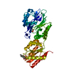


 Mass: 55.845 Da / Num. of mol.: 2 / Source method: isolated from a natural source / Formula: Fe
 Mass: 31.999 Da / Num. of mol.: 1 / Source method: obtained synthetically / Formula: O2
 Mass: 456.344 Da / Num. of mol.: 1 / Source method: obtained synthetically / Formula: C17H21N4O9P
 Mass: 18.015 Da / Num. of mol.: 128 / Source method: isolated from a natural source / Formula: H2O
 Sample preparation
 / Beamline: ID23-2 / Wavelength: 0.8726 Å
 Processing