 Movie
Movie Controller

[English] 日本語
 Yorodumi
- PDB-6frm: Crystal Structure of coenzyme F420H2 oxidase (FprA) co-crystalliz... -
+ Open data

- Basic information
| Entry | Database: PDB / ID: 6frm | ||||||
|---|---|---|---|---|---|---|---|
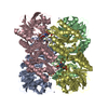
| Title | Crystal Structure of coenzyme F420H2 oxidase (FprA) co-crystallized with 10 mM Tb-Xo4 | ||||||
 Components Components | Coenzyme F420H2 oxidase (FprA) | ||||||
 Keywords Keywords | OXIDOREDUCTASE / Tb-Xo4 / crystallophore / FprA / nucleation / phasing / anomalous | ||||||
| Function / homology |  Function and homology information Function and homology informationFMN binding / electron transfer activity / oxidoreductase activity / metal ion binding Similarity search - Function | ||||||
| Biological species |  Methanothermococcus thermolithotrophicus (archaea) Methanothermococcus thermolithotrophicus (archaea) | ||||||
| Method |  X-RAY DIFFRACTION / SYNCHROTRON / MOLECULAR REPLACEMENT / Resolution: 2.2 Å X-RAY DIFFRACTION / SYNCHROTRON / MOLECULAR REPLACEMENT / Resolution: 2.2 Å | ||||||
 Authors Authors | Engilberge, S. / Riobe, F. / Wagner, T. / Di Pietro, S. / Shima, S. / Girard, E. / Dumont, E. / Maury, O. | ||||||
| Funding support |  France, 1items France, 1items
| ||||||
 Citation Citation | Journal: Chemistry / Year: 2018 Title: Unveiling the Binding Modes of the Crystallophore, a Terbium-based Nucleating and Phasing Molecular Agent for Protein Crystallography. Authors: Engilberge, S. / Riobe, F. / Wagner, T. / Di Pietro, S. / Breyton, C. / Franzetti, B. / Shima, S. / Girard, E. / Dumont, E. / Maury, O. | ||||||
| History |
|
- Structure visualization
| Structure viewer | Molecule: MolmilJmol/JSmol |
|---|
- Downloads & links
-Download
| PDBx/mmCIF format | 6frm.cif.gz | 1.3 MB | Display | PDBx/mmCIF format |
|---|---|---|---|---|
| PDB format | pdb6frm.ent.gz | 1.1 MB | Display | PDB format |
| PDBx/mmJSON format | 6frm.json.gz | Tree view | PDBx/mmJSON format | |
| Others |  Other downloads Other downloads |
-Validation report
| Arichive directory | https://data.pdbj.org/pub/pdb/validation_reports/fr/6frmftp://data.pdbj.org/pub/pdb/validation_reports/fr/6frm | HTTPS FTP |
|---|
-Related structure data
| Related structure data |  6f2fC  6f2hC 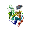 6f2iC 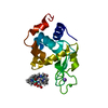 6f2jC  6f2kC  6f2mC 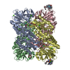 6frnC 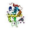 6froC  6frqC  2ohhS S: Starting model for refinement C: citing same article ( |
|---|---|
| Similar structure data |


-Links
 PDBj
PDBj


- Assembly
| Deposited unit | 
| ||||||||
|---|---|---|---|---|---|---|---|---|---|
| 1 | 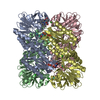
| ||||||||
| 2 | 
| ||||||||
| Unit cell |
|
-Components
-Protein , 1 types, 8 molecules ABCDEFGH
| #1: Protein | Mass: 46242.230 Da / Num. of mol.: 8 / Source method: isolated from a natural source Source: (natural) Methanothermococcus thermolithotrophicus (archaea)References: UniProt: A0A452CSW8*PLUS |
|---|
-Non-polymers , 6 types, 1626 molecules 


| #2: Chemical | ChemComp-FMN /  Mass: 456.344 Da / Num. of mol.: 8 / Source method: obtained synthetically / Formula: C17H21N4O9P Mass: 456.344 Da / Num. of mol.: 8 / Source method: obtained synthetically / Formula: C17H21N4O9P#3: Chemical |  Mass: 556.353 Da / Num. of mol.: 2 / Source method: obtained synthetically / Formula: C20H23N5O4Tb Mass: 556.353 Da / Num. of mol.: 2 / Source method: obtained synthetically / Formula: C20H23N5O4Tb#4: Chemical | ChemComp-TB /  Mass: 158.925 Da / Num. of mol.: 14 / Source method: obtained synthetically / Formula: Tb Mass: 158.925 Da / Num. of mol.: 14 / Source method: obtained synthetically / Formula: Tb#5: Chemical | ChemComp-CL /  Mass: 35.453 Da / Num. of mol.: 17 / Source method: obtained synthetically / Formula: Cl Mass: 35.453 Da / Num. of mol.: 17 / Source method: obtained synthetically / Formula: Cl#6: Chemical | ChemComp-FE /  Mass: 55.845 Da / Num. of mol.: 4 / Source method: obtained synthetically / Formula: Fe Mass: 55.845 Da / Num. of mol.: 4 / Source method: obtained synthetically / Formula: Fe#7: Water | ChemComp-HOH / | Mass: 18.015 Da / Num. of mol.: 1581 / Source method: isolated from a natural source / Formula: H2O |
|---|
-Experimental details
-Experiment
| Experiment | Method: X-RAY DIFFRACTION / Number of used crystals: 1 |
|---|
- Sample preparation
| Crystal | Density Matthews: 2.46 Å3/Da / Density % sol: 49.94 % |
|---|---|
| Crystal grow | Temperature: 291 K / Method: vapor diffusion, hanging drop Details: FprA native (about 90 % pure) at 43 g/l + 10 mM Xo4, crystallized at 291 K under anaerobic condition (95% N2 / 5% H2) in Combiclover Junior plate from Jena Bioscience. The protein was ...Details: FprA native (about 90 % pure) at 43 g/l + 10 mM Xo4, crystallized at 291 K under anaerobic condition (95% N2 / 5% H2) in Combiclover Junior plate from Jena Bioscience. The protein was crystallized by mixing 1 ul protein of this mix with 1 ul of the mother liquor containing 20% w/v PEG 8000, 100 mM HEPES pH 7.5, 200 mM Ammonium sulfate and 10 % v/v 2-propanol. Prior to freezing in liquid nitrogen, the crystal was soaked for few seconds in a cryo-protected solution containing: 20% w/v PEG 8000, 100 mM HEPES pH 7.5, 200 mM Ammonium sulfate, 10 % v/v 2-propanol, 25 % v/v glycerol. |
-Data collection
| Diffraction | Mean temperature: 100 K |
|---|---|
| Diffraction source | Source: SYNCHROTRON / Site: ESRF / Beamline: ID29 / Wavelength: 1.07227 Å |
| Detector | Type: DECTRIS PILATUS3 S 6M / Detector: PIXEL / Date: Aug 27, 2017 |
| Radiation | Protocol: SINGLE WAVELENGTH / Monochromatic (M) / Laue (L): M / Scattering type: x-ray |
| Radiation wavelength | Wavelength: 1.07227 Å / Relative weight: 1 |
| Reflection | Resolution: 2.19→48.69 Å / Num. obs: 181493 / % possible obs: 99 % / Redundancy: 6.7 % / Biso Wilson estimate: 44.22 Å2 / Rpim(I) all: 0.044 / Net I/σ(I): 9 |
| Reflection shell | Resolution: 2.19→2.31 Å / Rpim(I) all: 0.534 |
- Processing
| Software |
| ||||||||||||||||||||||||||||||||||||||||||||||||||||||||||||||||||||||||||||||||||||||||||||||||||||||||||||||||||||||||||||||||||||||||||||||||||||||||||||||||||||||||||||||||||||||||||||||||||||||||||||||||||||||||||||||||||||||||||||||||||||||||||
|---|---|---|---|---|---|---|---|---|---|---|---|---|---|---|---|---|---|---|---|---|---|---|---|---|---|---|---|---|---|---|---|---|---|---|---|---|---|---|---|---|---|---|---|---|---|---|---|---|---|---|---|---|---|---|---|---|---|---|---|---|---|---|---|---|---|---|---|---|---|---|---|---|---|---|---|---|---|---|---|---|---|---|---|---|---|---|---|---|---|---|---|---|---|---|---|---|---|---|---|---|---|---|---|---|---|---|---|---|---|---|---|---|---|---|---|---|---|---|---|---|---|---|---|---|---|---|---|---|---|---|---|---|---|---|---|---|---|---|---|---|---|---|---|---|---|---|---|---|---|---|---|---|---|---|---|---|---|---|---|---|---|---|---|---|---|---|---|---|---|---|---|---|---|---|---|---|---|---|---|---|---|---|---|---|---|---|---|---|---|---|---|---|---|---|---|---|---|---|---|---|---|---|---|---|---|---|---|---|---|---|---|---|---|---|---|---|---|---|---|---|---|---|---|---|---|---|---|---|---|---|---|---|---|---|---|---|---|---|---|---|---|---|---|---|---|---|---|---|---|---|---|
| Refinement | Method to determine structure: MOLECULAR REPLACEMENT Starting model: 2OHH Resolution: 2.2→48.69 Å / Cor.coef. Fo:Fc: 0.956 / Cor.coef. Fo:Fc free: 0.937 / SU R Cruickshank DPI: 0.247 / Cross valid method: THROUGHOUT / σ(F): 0 / SU R Blow DPI: 0.247 / SU Rfree Blow DPI: 0.179 / SU Rfree Cruickshank DPI: 0.181
| ||||||||||||||||||||||||||||||||||||||||||||||||||||||||||||||||||||||||||||||||||||||||||||||||||||||||||||||||||||||||||||||||||||||||||||||||||||||||||||||||||||||||||||||||||||||||||||||||||||||||||||||||||||||||||||||||||||||||||||||||||||||||||
| Displacement parameters | Biso mean: 52.27 Å2
| ||||||||||||||||||||||||||||||||||||||||||||||||||||||||||||||||||||||||||||||||||||||||||||||||||||||||||||||||||||||||||||||||||||||||||||||||||||||||||||||||||||||||||||||||||||||||||||||||||||||||||||||||||||||||||||||||||||||||||||||||||||||||||
| Refine analyze | Luzzati coordinate error obs: 0.28 Å | ||||||||||||||||||||||||||||||||||||||||||||||||||||||||||||||||||||||||||||||||||||||||||||||||||||||||||||||||||||||||||||||||||||||||||||||||||||||||||||||||||||||||||||||||||||||||||||||||||||||||||||||||||||||||||||||||||||||||||||||||||||||||||
| Refinement step | Cycle: 1 / Resolution: 2.2→48.69 Å
| ||||||||||||||||||||||||||||||||||||||||||||||||||||||||||||||||||||||||||||||||||||||||||||||||||||||||||||||||||||||||||||||||||||||||||||||||||||||||||||||||||||||||||||||||||||||||||||||||||||||||||||||||||||||||||||||||||||||||||||||||||||||||||
| Refine LS restraints |
| ||||||||||||||||||||||||||||||||||||||||||||||||||||||||||||||||||||||||||||||||||||||||||||||||||||||||||||||||||||||||||||||||||||||||||||||||||||||||||||||||||||||||||||||||||||||||||||||||||||||||||||||||||||||||||||||||||||||||||||||||||||||||||
| LS refinement shell | Resolution: 2.2→2.26 Å / Total num. of bins used: 20
| ||||||||||||||||||||||||||||||||||||||||||||||||||||||||||||||||||||||||||||||||||||||||||||||||||||||||||||||||||||||||||||||||||||||||||||||||||||||||||||||||||||||||||||||||||||||||||||||||||||||||||||||||||||||||||||||||||||||||||||||||||||||||||
| Refinement TLS params. | Method: refined / Refine-ID: X-RAY DIFFRACTION
| ||||||||||||||||||||||||||||||||||||||||||||||||||||||||||||||||||||||||||||||||||||||||||||||||||||||||||||||||||||||||||||||||||||||||||||||||||||||||||||||||||||||||||||||||||||||||||||||||||||||||||||||||||||||||||||||||||||||||||||||||||||||||||
| Refinement TLS group |
|
