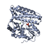
 Movie
Movie Controller


+ Open data

- Basic information
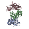
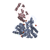
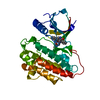
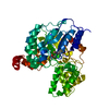
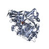
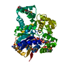
| Entry | Database: PDB / ID: 2y1n | ||||||
|---|---|---|---|---|---|---|---|
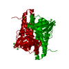
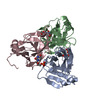
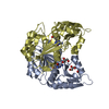
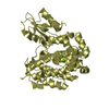
| Title | Structure of c-Cbl-ZAP-70 peptide complex | ||||||
 Components Components |
| ||||||
 Keywords Keywords | LIGASE/TRANSFERASE / LIGASE-TRANSFERASE COMPLEX / UBIQUITIN RING E3 LIGASE | ||||||
| Function / homology |  Function and homology information Function and homology informationT cell aggregation / ubiquitin-dependent endocytosis / regulation of platelet-derived growth factor receptor-alpha signaling pathway / T cell migration / regulation of Rap protein signal transduction / entry of bacterium into host cell / positive regulation of alpha-beta T cell proliferation / negative thymic T cell selection / negative regulation of epidermal growth factor receptor signaling pathway / flotillin complex ...T cell aggregation / ubiquitin-dependent endocytosis / regulation of platelet-derived growth factor receptor-alpha signaling pathway / T cell migration / regulation of Rap protein signal transduction / entry of bacterium into host cell / positive regulation of alpha-beta T cell proliferation / negative thymic T cell selection / negative regulation of epidermal growth factor receptor signaling pathway / flotillin complex / phosphatidylinositol 3-kinase regulatory subunit binding / beta selection / positive thymic T cell selection / positive regulation of alpha-beta T cell differentiation / positive regulation of T cell differentiation / positive regulation of epidermal growth factor receptor signaling pathway / T cell receptor complex / Regulation of KIT signaling / mast cell degranulation / Interleukin-6 signaling / response to testosterone / B cell activation / cellular response to platelet-derived growth factor stimulus / response to starvation / Translocation of ZAP-70 to Immunological synapse / protein monoubiquitination / TGF-beta receptor signaling activates SMADs / extrinsic component of cytoplasmic side of plasma membrane / RHOH GTPase cycle / Generation of second messenger molecules / immunological synapse / T cell differentiation / protein autoubiquitination / Nuclear events stimulated by ALK signaling in cancer / cell surface receptor protein tyrosine kinase signaling pathway / FLT3 signaling by CBL mutants / PTK6 Regulates RTKs and Their Effectors AKT1 and DOK1 / Negative regulation of FLT3 / T cell activation / positive regulation of calcium-mediated signaling / ephrin receptor binding / phosphotyrosine residue binding / EGFR downregulation / InlB-mediated entry of Listeria monocytogenes into host cell / cellular response to nerve growth factor stimulus / Constitutive Signaling by EGFRvIII / response to activity / Regulation of signaling by CBL / response to gamma radiation / Negative regulation of FGFR3 signaling / Negative regulation of FGFR2 signaling / Negative regulation of FGFR4 signaling / Negative regulation of FGFR1 signaling / non-specific protein-tyrosine kinase / calcium-mediated signaling / Spry regulation of FGF signaling / non-membrane spanning protein tyrosine kinase activity / RING-type E3 ubiquitin transferase / peptidyl-tyrosine phosphorylation / Negative regulation of MET activity / cilium / receptor tyrosine kinase binding / cytokine-mediated signaling pathway / positive regulation of receptor-mediated endocytosis / SH3 domain binding / protein polyubiquitination / Signaling by CSF1 (M-CSF) in myeloid cells / ubiquitin-protein transferase activity / Cargo recognition for clathrin-mediated endocytosis / male gonad development / ubiquitin protein ligase activity / Constitutive Signaling by Ligand-Responsive EGFR Cancer Variants / cell-cell junction / Clathrin-mediated endocytosis / cellular response to hypoxia / T cell receptor signaling pathway / protein tyrosine kinase activity / growth cone / ubiquitin-dependent protein catabolic process / response to ethanol / adaptive immune response / positive regulation of phosphatidylinositol 3-kinase/protein kinase B signal transduction / cell differentiation / intracellular signal transduction / protein ubiquitination / cadherin binding / membrane raft / immune response / protein phosphorylation / innate immune response / focal adhesion / signaling receptor binding / DNA damage response / calcium ion binding / negative regulation of apoptotic process / perinuclear region of cytoplasm / Golgi apparatus / signal transduction / ATP binding / plasma membrane Similarity search - Function | ||||||
| Biological species |  HOMO SAPIENS (human) HOMO SAPIENS (human) | ||||||
| Method |  X-RAY DIFFRACTION / SYNCHROTRON / MOLECULAR REPLACEMENT / Resolution: 1.999 Å X-RAY DIFFRACTION / SYNCHROTRON / MOLECULAR REPLACEMENT / Resolution: 1.999 Å | ||||||
 Authors Authors | Dou, H. / Sibbet, G.J. / Huang, D.T. | ||||||
 Citation Citation | Journal: Nat.Struct.Mol.Biol. / Year: 2012 Title: Structural Basis for Autoinhibition and Phosphorylation-Dependent Activation of C-Cbl. Authors: Dou, H. / Buetow, L. / Hock, A. / Sibbet, G.J. / Vousden, K.H. / Huang, D.T. | ||||||
| History |
|
- Structure visualization
| Structure viewer | Molecule: MolmilJmol/JSmol |
|---|
- Downloads & links
-Download
| PDBx/mmCIF format | 2y1n.cif.gz | 175.5 KB | Display | PDBx/mmCIF format |
|---|---|---|---|---|
| PDB format | pdb2y1n.ent.gz | 138.5 KB | Display | PDB format |
| PDBx/mmJSON format | 2y1n.json.gz | Tree view | PDBx/mmJSON format | |
| Others |  Other downloads Other downloads |
-Validation report
| Summary document | 2y1n_validation.pdf.gz | 460.7 KB | Display | wwPDB validaton report |
|---|---|---|---|---|
| Full document | 2y1n_full_validation.pdf.gz | 474.4 KB | Display | |
| Data in XML | 2y1n_validation.xml.gz | 33.6 KB | Display | |
| Data in CIF | 2y1n_validation.cif.gz | 47.6 KB | Display | |
| Arichive directory | https://data.pdbj.org/pub/pdb/validation_reports/y1/2y1nftp://data.pdbj.org/pub/pdb/validation_reports/y1/2y1n | HTTPS FTP |
-Related structure data
| Related structure data | 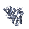 2y1mC 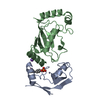 4a49C 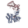 4a4bC 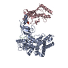 4a4cC 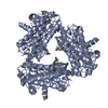 2cblS S: Starting model for refinement C: citing same article ( |
|---|---|
| Similar structure data |


-Links
 PDBj
PDBj


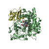
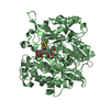
- Assembly
| Deposited unit | 
| ||||||||
|---|---|---|---|---|---|---|---|---|---|
| 1 | 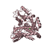
| ||||||||
| 2 | 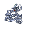
| ||||||||
| Unit cell |
|
-Components
| #1: Protein | Mass: 44928.695 Da / Num. of mol.: 2 / Fragment: RESIDUES 47-435 Source method: isolated from a genetically manipulated source Source: (gene. exp.) HOMO SAPIENS (human) / Production host:  References: UniProt: P22681, Ligases; Forming carbon-nitrogen bonds; Acid-amino-acid ligases (peptide synthases) #2: Protein/peptide | Mass: 1344.275 Da / Num. of mol.: 2 / Fragment: PEPTIDE, RESIDUES 286-297 / Source method: obtained synthetically Details: ZAP-70 PEPTIDE (TLNSDG(P)YTPEPA) WITH PHOSPHOTYROSINE AT POSITION 7 Source: (synth.) HOMO SAPIENS (human)References: UniProt: P43403, non-specific protein-tyrosine kinase #3: Chemical | ChemComp-ZN /   Mass: 65.409 Da / Num. of mol.: 4 / Source method: obtained synthetically / Formula: Zn Mass: 65.409 Da / Num. of mol.: 4 / Source method: obtained synthetically / Formula: Zn#4: Chemical |   Mass: 40.078 Da / Num. of mol.: 2 / Source method: obtained synthetically / Formula: Ca Mass: 40.078 Da / Num. of mol.: 2 / Source method: obtained synthetically / Formula: Ca#5: Water | ChemComp-HOH / |  Mass: 18.015 Da / Num. of mol.: 340 / Source method: isolated from a natural source / Formula: H2O Mass: 18.015 Da / Num. of mol.: 340 / Source method: isolated from a natural source / Formula: H2O |
|---|
-Experimental details
-Experiment
| Experiment | Method: X-RAY DIFFRACTION / Number of used crystals: 1 |
|---|
- Sample preparation
| Crystal | Density Matthews: 2.66 Å3/Da / Density % sol: 53.83 % / Description: NONE |
|---|---|
| Crystal grow | Details: 18-20% (W/V) PEG 3350, 0.2 M AMMONIUM FORMATE |
-Data collection
| Diffraction | Mean temperature: 100 K |
|---|---|
| Diffraction source | Source: SYNCHROTRON / Site: Diamond  / Beamline: I03 / Wavelength: 0.97625 / Beamline: I03 / Wavelength: 0.97625 |
| Detector | Type: ADSC CCD / Detector: CCD / Date: Jul 8, 2010 |
| Radiation | Monochromator: SYNCHROTRON / Protocol: SINGLE WAVELENGTH / Monochromatic (M) / Laue (L): M / Scattering type: x-ray |
| Radiation wavelength | Wavelength: 0.97625 Å / Relative weight: 1 |
| Reflection twin | Operator: h,-h-k,-l / Fraction: 0.395 |
| Reflection | Resolution: 2→50 Å / Num. obs: 63238 / % possible obs: 99.9 % / Observed criterion σ(I): 2 / Redundancy: 3.8 % / Biso Wilson estimate: 33.2 Å2 / Rmerge(I) obs: 0.08 / Net I/σ(I): 30.3 |
| Reflection shell | Resolution: 2→2.07 Å / Redundancy: 3.4 % / Rmerge(I) obs: 0.52 / Mean I/σ(I) obs: 2.5 / % possible all: 99.7 |
- Processing
| Software |
| |||||||||||||||||||||||||||||||||||||||||||||||||||||||||||||||||||||||||||||||||||||||||||||||||||||||||||||||||||||||||||||||||||||||||||||||||||
|---|---|---|---|---|---|---|---|---|---|---|---|---|---|---|---|---|---|---|---|---|---|---|---|---|---|---|---|---|---|---|---|---|---|---|---|---|---|---|---|---|---|---|---|---|---|---|---|---|---|---|---|---|---|---|---|---|---|---|---|---|---|---|---|---|---|---|---|---|---|---|---|---|---|---|---|---|---|---|---|---|---|---|---|---|---|---|---|---|---|---|---|---|---|---|---|---|---|---|---|---|---|---|---|---|---|---|---|---|---|---|---|---|---|---|---|---|---|---|---|---|---|---|---|---|---|---|---|---|---|---|---|---|---|---|---|---|---|---|---|---|---|---|---|---|---|---|---|---|
| Refinement | Method to determine structure: MOLECULAR REPLACEMENT Starting model: PDB ENTRY 2CBL Resolution: 1.999→45.423 Å / σ(F): 1.36 / Phase error: 30.1 / Stereochemistry target values: TWIN_LSQ_F Details: RESIDUES OMITTED DUE TO POORLY DEFINED ELECTRON DENSITY. CHAIN A RESIDUES 355-361, CHAIN C RESIDUES 355-35 CHAINS B AND D RESIDUES 1-3. DUE TO POORLY DEFINED ELECTRON DENSITY, LYS53 AND ...Details: RESIDUES OMITTED DUE TO POORLY DEFINED ELECTRON DENSITY. CHAIN A RESIDUES 355-361, CHAIN C RESIDUES 355-35 CHAINS B AND D RESIDUES 1-3. DUE TO POORLY DEFINED ELECTRON DENSITY, LYS53 AND LYS54 IN CHAINS A AND C WERE BUILT AS ALANINES.
| |||||||||||||||||||||||||||||||||||||||||||||||||||||||||||||||||||||||||||||||||||||||||||||||||||||||||||||||||||||||||||||||||||||||||||||||||||
| Solvent computation | Shrinkage radii: 0.9 Å / VDW probe radii: 1.11 Å / Solvent model: FLAT BULK SOLVENT MODEL / Bsol: 30.127 Å2 / ksol: 0.327 e/Å3 | |||||||||||||||||||||||||||||||||||||||||||||||||||||||||||||||||||||||||||||||||||||||||||||||||||||||||||||||||||||||||||||||||||||||||||||||||||
| Displacement parameters | Biso mean: 39.82 Å2
| |||||||||||||||||||||||||||||||||||||||||||||||||||||||||||||||||||||||||||||||||||||||||||||||||||||||||||||||||||||||||||||||||||||||||||||||||||
| Refinement step | Cycle: LAST / Resolution: 1.999→45.423 Å
| |||||||||||||||||||||||||||||||||||||||||||||||||||||||||||||||||||||||||||||||||||||||||||||||||||||||||||||||||||||||||||||||||||||||||||||||||||
| Refine LS restraints |
| |||||||||||||||||||||||||||||||||||||||||||||||||||||||||||||||||||||||||||||||||||||||||||||||||||||||||||||||||||||||||||||||||||||||||||||||||||
| LS refinement shell |
|
