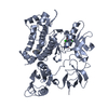
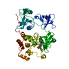
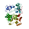
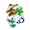
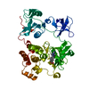
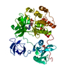
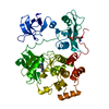
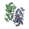
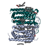
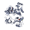
Entry Database : PDB / ID : 2fo0Title Organization of the SH3-SH2 Unit in Active and Inactive Forms of the c-Abl Tyrosine Kinase Proto-oncogene tyrosine-protein kinase ABL1 (1B ISOFORM) Keywords / / / / / Function / homology Function Domain/homology Component
/ / / / / / / / / / / / / / / / / / / / / / / / / / / / / / / / / / / / / / / / / / / / / / / / / / / / / / / / / / / / / / / / / / / / / / / / / / / / / / / / / / / / / / / / / / / / / / / / / / / / / / / / / / / / / / / / / / / / / / / / / / / / / / / / / / / / / / / / / / / / / / / / / / / / / / / / / / / / Biological species Homo sapiens (human)Method / / / Resolution : 2.27 Å Authors Nagar, B. / Hantschel, O. / Seeliger, M. / Davies, J.M. / Weis, W.I. / Superti-Furga, G. / Kuriyan, J. Journal : Mol.Cell / Year : 2006Title : Organization of the SH3-SH2 unit in active and inactive forms of the c-Abl tyrosine kinase.Authors : Nagar, B. / Hantschel, O. / Seeliger, M. / Davies, J.M. / Weis, W.I. / Superti-Furga, G. / Kuriyan, J. History Deposition Jan 12, 2006 Deposition site / Processing site Revision 1.0 Mar 21, 2006 Provider / Type Revision 1.1 May 1, 2008 Group Revision 1.2 Jul 13, 2011 Group / Version format complianceRevision 1.3 Aug 2, 2017 Group / Category Revision 2.0 Jun 30, 2021 Group Advisory / Atomic model ... Advisory / Atomic model / Data collection / Database references / Derived calculations Category atom_site / pdbx_nonpoly_scheme ... atom_site / pdbx_nonpoly_scheme / pdbx_unobs_or_zero_occ_atoms / struct_conn / struct_ref_seq_dif / struct_site Item _atom_site.auth_seq_id / _pdbx_nonpoly_scheme.pdb_seq_num ... _atom_site.auth_seq_id / _pdbx_nonpoly_scheme.pdb_seq_num / _pdbx_unobs_or_zero_occ_atoms.auth_seq_id / _struct_conn.pdbx_leaving_atom_flag / _struct_ref_seq_dif.details / _struct_site.details / _struct_site.pdbx_auth_asym_id / _struct_site.pdbx_auth_comp_id / _struct_site.pdbx_auth_seq_id Revision 2.1 Oct 20, 2021 Group / Category / struct_ref_seq_difItem / _database_2.pdbx_database_accession / _struct_ref_seq_dif.detailsRevision 2.2 Aug 30, 2023 Group / Refinement descriptionCategory / chem_comp_bond / pdbx_initial_refinement_modelRevision 2.3 Oct 30, 2024 Group / Category / pdbx_modification_feature
Show all Show less Remark 999 SEQUENCE Residues 15-56 of the original protein sequence were deleted. Myristoyl group (MYR) is ... SEQUENCE Residues 15-56 of the original protein sequence were deleted. Myristoyl group (MYR) is covalently attached to the N-terminus of the protein.
 Movie
Movie Controller

 Yorodumi
 Open data
 Basic information
 Components
 Keywords
 Function and homology information
 Homo sapiens (human)
 X-RAY DIFFRACTION /
  Authors
 Citation
 Structure visualization
 Downloads & links
 Other downloads
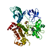
 PDBj
PDBj


 Assembly
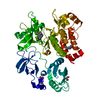

 Spodoptera frugiperda (fall armyworm)
 Mass: 228.371 Da / Num. of mol.: 1 / Source method: obtained synthetically / Formula: C14H28O2
 Mass: 427.283 Da / Num. of mol.: 1 / Source method: obtained synthetically / Formula: C21H16Cl2N4O2
 Mass: 92.094 Da / Num. of mol.: 2 / Source method: obtained synthetically / Formula: C3H8O3
 Mass: 18.015 Da / Num. of mol.: 145 / Source method: isolated from a natural source / Formula: H2O
 Sample preparation
 / Beamline: 8.2.2 / Wavelength: 1.1 Å
 Processing