 Movie
Movie Controller

[English] 日本語
 Yorodumi
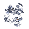
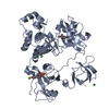
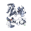
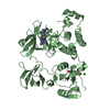
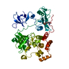
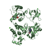
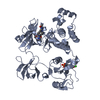
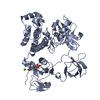
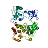
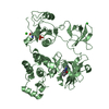
- PDB-1qcf: CRYSTAL STRUCTURE OF HCK IN COMPLEX WITH A SRC FAMILY-SELECTIVE T... -
+ Open data

- Basic information
| Entry | Database: PDB / ID: 1qcf | ||||||
|---|---|---|---|---|---|---|---|
| Title | CRYSTAL STRUCTURE OF HCK IN COMPLEX WITH A SRC FAMILY-SELECTIVE TYROSINE KINASE INHIBITOR | ||||||
 Components Components | Tyrosine-protein kinase HCK | ||||||
 Keywords Keywords | TYROSINE KINASE / TYROSINE KINASE-INHIBITOR COMPLEX / DOWN-REGULATED KINASE / ORDERED ACTIVATION LOOP | ||||||
| Function / homology |  Function and homology information Function and homology informationleukocyte degranulation / leukocyte migration involved in immune response / respiratory burst after phagocytosis / regulation of podosome assembly / innate immune response-activating signaling pathway / : / FLT3 signaling through SRC family kinases / regulation of phagocytosis / Nef and signal transduction / Fc-gamma receptor signaling pathway involved in phagocytosis ...leukocyte degranulation / leukocyte migration involved in immune response / respiratory burst after phagocytosis / regulation of podosome assembly / innate immune response-activating signaling pathway / : / FLT3 signaling through SRC family kinases / regulation of phagocytosis / Nef and signal transduction / Fc-gamma receptor signaling pathway involved in phagocytosis / mesoderm development / positive regulation of actin filament polymerization / FCGR activation / type II interferon-mediated signaling pathway / transport vesicle / Signaling by CSF3 (G-CSF) / cell projection / phosphotyrosine residue binding / peptidyl-tyrosine phosphorylation / lipopolysaccharide-mediated signaling pathway / FCGR3A-mediated IL10 synthesis / cell surface receptor protein tyrosine kinase signaling pathway / integrin-mediated signaling pathway / regulation of actin cytoskeleton organization / non-specific protein-tyrosine kinase / FCGR3A-mediated phagocytosis / Regulation of signaling by CBL / negative regulation of inflammatory response to antigenic stimulus / non-membrane spanning protein tyrosine kinase activity / caveola / Inactivation of CSF3 (G-CSF) signaling / cytoplasmic side of plasma membrane / cytokine-mediated signaling pathway / Signaling by CSF1 (M-CSF) in myeloid cells / protein autophosphorylation / regulation of cell shape / protein tyrosine kinase activity / regulation of inflammatory response / cytoskeleton / protein phosphorylation / cell differentiation / lysosome / cell adhesion / intracellular signal transduction / defense response to Gram-positive bacterium / inflammatory response / signaling receptor binding / focal adhesion / positive regulation of cell population proliferation / lipid binding / negative regulation of apoptotic process / Golgi apparatus / ATP binding / nucleus / plasma membrane / cytosol Similarity search - Function | ||||||
| Biological species |  Homo sapiens (human) Homo sapiens (human) | ||||||
| Method |  X-RAY DIFFRACTION / Resolution: 2 Å X-RAY DIFFRACTION / Resolution: 2 Å | ||||||
 Authors Authors | Schindler, T. / Sicheri, F. / Pico, A. / Gazit, A. / Levitzki, A. / Kuriyan, J. | ||||||
 Citation Citation | Journal: Mol.Cell / Year: 1999 Title: Crystal structure of Hck in complex with a Src family-selective tyrosine kinase inhibitor. Authors: Schindler, T. / Sicheri, F. / Pico, A. / Gazit, A. / Levitzki, A. / Kuriyan, J. | ||||||
| History |
|
- Structure visualization
| Structure viewer | Molecule: MolmilJmol/JSmol |
|---|
- Downloads & links
-Download
| PDBx/mmCIF format | 1qcf.cif.gz | 112.2 KB | Display | PDBx/mmCIF format |
|---|---|---|---|---|
| PDB format | pdb1qcf.ent.gz | 84.1 KB | Display | PDB format |
| PDBx/mmJSON format | 1qcf.json.gz | Tree view | PDBx/mmJSON format | |
| Others |  Other downloads Other downloads |
-Validation report
| Arichive directory | https://data.pdbj.org/pub/pdb/validation_reports/qc/1qcfftp://data.pdbj.org/pub/pdb/validation_reports/qc/1qcf | HTTPS FTP |
|---|
-Related structure data
| Related structure data | |
|---|---|
| Similar structure data |

-Links
 PDBj
PDBj


- Assembly
| Deposited unit | 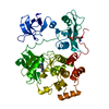
| ||||||||
|---|---|---|---|---|---|---|---|---|---|
| 1 |
| ||||||||
| Unit cell |
|
-Components
| #1: Protein | Mass: 52000.227 Da / Num. of mol.: 1 / Fragment: SH3-SH2-KINASE-HIGH AFFINITY TAIL / Mutation: Q528E, Q529E, Q530I Source method: isolated from a genetically manipulated source Source: (gene. exp.) Homo sapiens (human) / Gene: HCKReferences: UniProt: P08631, non-specific protein-tyrosine kinase |
|---|---|
| #2: Chemical | ChemComp-PP1 /   Mass: 281.356 Da / Num. of mol.: 1 / Source method: obtained synthetically / Formula: C16H19N5 Mass: 281.356 Da / Num. of mol.: 1 / Source method: obtained synthetically / Formula: C16H19N5 |
| #3: Water | ChemComp-HOH /  Mass: 18.015 Da / Num. of mol.: 312 / Source method: isolated from a natural source / Formula: H2O Mass: 18.015 Da / Num. of mol.: 312 / Source method: isolated from a natural source / Formula: H2O |
| Has protein modification | Y |
-Experimental details
-Experiment
| Experiment | Method: X-RAY DIFFRACTION / Number of used crystals: 1 |
|---|
- Sample preparation
| Crystal | Density Matthews: 2.51 Å3/Da / Density % sol: 50.98 % | |||||||||||||||||||||||||
|---|---|---|---|---|---|---|---|---|---|---|---|---|---|---|---|---|---|---|---|---|---|---|---|---|---|---|
| Crystal grow | Temperature: 293 K / Method: vapor diffusion, hanging drop / pH: 7 Details: PEG 10000, DIMETHYL SULFOXIDE, N-(2-HYDROXYETHYL)PIPERAZINE-N-(2- ETHANESULFONIC ACID), pH 7.0, VAPOR DIFFUSION, HANGING DROP, temperature 293K | |||||||||||||||||||||||||
| Crystal grow | *PLUS Details: drop solution was mixed with an equal volume of reservoir solution | |||||||||||||||||||||||||
| Components of the solutions | *PLUS
|
-Data collection
| Diffraction | Mean temperature: 105 K |
|---|---|
| Diffraction source | Source: ROTATING ANODE / Type: RIGAKU RU200 / Wavelength: 1.5418 |
| Detector | Type: RIGAKU RAXIS IIC / Detector: IMAGE PLATE / Date: Oct 22, 1999 |
| Radiation | Protocol: SINGLE WAVELENGTH / Monochromatic (M) / Laue (L): M / Scattering type: x-ray |
| Radiation wavelength | Wavelength: 1.5418 Å / Relative weight: 1 |
| Reflection | Resolution: 2→99 Å / Num. all: 35649 / Num. obs: 35042 / % possible obs: 98.3 % / Observed criterion σ(F): 0 / Observed criterion σ(I): 0 / Redundancy: 6.4 % / Biso Wilson estimate: 25.6 Å2 / Rmerge(I) obs: 0.086 / Net I/σ(I): 28.2 |
| Reflection shell | Resolution: 2→2.1 Å / Redundancy: 5.4 % / Rmerge(I) obs: 0.277 / % possible all: 95.2 |
| Reflection | *PLUS Num. measured all: 426133 |
| Reflection shell | *PLUS % possible obs: 95.2 % / Num. unique obs: 4250 / Num. measured obs: 23990 |
- Processing
| Software |
| ||||||||||||||||||||||||||||||||||||||||||||||||||||||||||||
|---|---|---|---|---|---|---|---|---|---|---|---|---|---|---|---|---|---|---|---|---|---|---|---|---|---|---|---|---|---|---|---|---|---|---|---|---|---|---|---|---|---|---|---|---|---|---|---|---|---|---|---|---|---|---|---|---|---|---|---|---|---|
| Refinement | Resolution: 2→99 Å / σ(F): 0 / σ(I): 0 / Stereochemistry target values: ENGH & HUBER
| ||||||||||||||||||||||||||||||||||||||||||||||||||||||||||||
| Refinement step | Cycle: LAST / Resolution: 2→99 Å
| ||||||||||||||||||||||||||||||||||||||||||||||||||||||||||||
| Refine LS restraints |
| ||||||||||||||||||||||||||||||||||||||||||||||||||||||||||||
| Software | *PLUS Name: 'CNS' / Classification: refinement | ||||||||||||||||||||||||||||||||||||||||||||||||||||||||||||
| Refinement | *PLUS Rfactor obs: 0.215 | ||||||||||||||||||||||||||||||||||||||||||||||||||||||||||||
| Solvent computation | *PLUS | ||||||||||||||||||||||||||||||||||||||||||||||||||||||||||||
| Displacement parameters | *PLUS | ||||||||||||||||||||||||||||||||||||||||||||||||||||||||||||
| Refine LS restraints | *PLUS
|
