 Movie
Movie Controller

[English] 日本語
 Yorodumi
- PDB-2eh8: Crystal structure of the complex of humanized KR127 fab and PRES1... -
+ Open data

- Basic information
| Entry | Database: PDB / ID: 2eh8 | ||||||
|---|---|---|---|---|---|---|---|
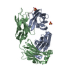
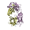
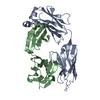
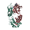
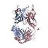
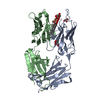
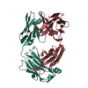
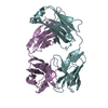
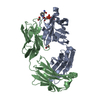
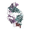
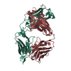
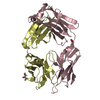
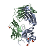
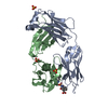
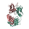
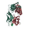
| Title | Crystal structure of the complex of humanized KR127 fab and PRES1 peptide epitope | ||||||
 Components Components |
| ||||||
 Keywords Keywords | IMMUNE SYSTEM / HEPATITIS B VIRUS / HUMANIZED ANTIBODY / MONOCLONAL ANTIBODY / NEUTRALIZATION / PRES1 | ||||||
| Function / homology |  Function and homology information Function and homology informationcaveolin-mediated endocytosis of virus by host cell / membrane => GO:0016020 / fusion of virus membrane with host endosome membrane / virion attachment to host cell / virion membrane Similarity search - Function | ||||||
| Biological species |  | ||||||
| Method |  X-RAY DIFFRACTION / MOLECULAR REPLACEMENT / Resolution: 2.6 Å X-RAY DIFFRACTION / MOLECULAR REPLACEMENT / Resolution: 2.6 Å | ||||||
 Authors Authors | Chi, S.-W. / Kim, S.-J. / Maeng, C.-Y. / Hong, H.J. / Ryu, S.-E. | ||||||
 Citation Citation | Journal: Proc.Natl.Acad.Sci.Usa / Year: 2007 Title: Broadly neutralizing anti-hepatitis B virus antibody reveals a complementarity determining region H3 lid-opening mechanism Authors: Chi, S.-W. / Maeng, C.-Y. / Kim, S.J. / Oh, M.S. / Ryu, C.J. / Kim, S.-J. / Han, K.-H. / Hong, H.J. / Ryu, S.-E. | ||||||
| History |
|
- Structure visualization
| Structure viewer | Molecule: MolmilJmol/JSmol |
|---|
- Downloads & links
-Download
| PDBx/mmCIF format | 2eh8.cif.gz | 98.3 KB | Display | PDBx/mmCIF format |
|---|---|---|---|---|
| PDB format | pdb2eh8.ent.gz | 73.9 KB | Display | PDB format |
| PDBx/mmJSON format | 2eh8.json.gz | Tree view | PDBx/mmJSON format | |
| Others |  Other downloads Other downloads |
-Validation report
| Summary document | 2eh8_validation.pdf.gz | 429.9 KB | Display | wwPDB validaton report |
|---|---|---|---|---|
| Full document | 2eh8_full_validation.pdf.gz | 437.7 KB | Display | |
| Data in XML | 2eh8_validation.xml.gz | 18.3 KB | Display | |
| Data in CIF | 2eh8_validation.cif.gz | 24.6 KB | Display | |
| Arichive directory | https://data.pdbj.org/pub/pdb/validation_reports/eh/2eh8ftp://data.pdbj.org/pub/pdb/validation_reports/eh/2eh8 | HTTPS FTP |
-Related structure data
| Related structure data | 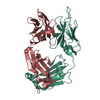 2eh7SC S: Starting model for refinement C: citing same article ( |
|---|---|
| Similar structure data |


-Links
 PDBj
PDBj

- Assembly
| Deposited unit | 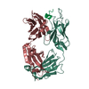
| ||||||||
|---|---|---|---|---|---|---|---|---|---|
| 1 |
| ||||||||
| Unit cell |
|
-Components
| #1: Antibody | Mass: 23788.590 Da / Num. of mol.: 1 Source method: isolated from a genetically manipulated source Source: (gene. exp.)  Homo sapiens (human) Homo sapiens (human) |
|---|---|
| #2: Antibody | Mass: 23086.756 Da / Num. of mol.: 1 Source method: isolated from a genetically manipulated source Source: (gene. exp.) Homo sapiens (human) |
| #3: Protein/peptide | Mass: 1293.256 Da / Num. of mol.: 1 / Source method: obtained synthetically Details: The peptide P was chemically synthesized. The sequence is naturally found in hepatitis B virus. References: UniProt: Q2EID8 |
| #4: Water | ChemComp-HOH /  Mass: 18.015 Da / Num. of mol.: 41 / Source method: isolated from a natural source / Formula: H2O Mass: 18.015 Da / Num. of mol.: 41 / Source method: isolated from a natural source / Formula: H2O |
| Sequence details | THE SHORT PEPTIDE, PRES1, IS AMIDATED /ACETYLATED AT THE N- AND C-TERMINUS, RESPECTIVELY. A ...THE SHORT PEPTIDE, PRES1, IS AMIDATED /ACETYLATED |
-Experimental details
-Experiment
| Experiment | Method: X-RAY DIFFRACTION / Number of used crystals: 1 |
|---|
- Sample preparation
| Crystal | Density Matthews: 2.53 Å3/Da / Density % sol: 51.36 % |
|---|---|
| Crystal grow | Temperature: 288 K / Method: vapor diffusion, hanging drop / pH: 7.5 Details: 17% PEG 4000, 10mM Hepes-NaOH, 0.2M ammonium sulfate, pH 7.50, VAPOR DIFFUSION, HANGING DROP, temperature 288K |
-Data collection
| Diffraction | Mean temperature: 298 K |
|---|---|
| Diffraction source | Source: ROTATING ANODE / Type: RIGAKU RU300 / Wavelength: 1.5418 / Wavelength: 1.5418 Å |
| Detector | Type: RIGAKU RAXIS IV / Detector: IMAGE PLATE / Date: Jul 30, 2001 |
| Radiation | Monochromator: CONFOCAL MIRROR / Protocol: SINGLE WAVELENGTH / Monochromatic (M) / Laue (L): M / Scattering type: x-ray |
| Radiation wavelength | Wavelength: 1.5418 Å / Relative weight: 1 |
| Reflection | Resolution: 2.6→32.91 Å / Num. all: 14984 / Num. obs: 14611 / % possible obs: 100 % / Observed criterion σ(F): 0 / Observed criterion σ(I): 0 / Redundancy: 3.6 % / Rmerge(I) obs: 0.075 / Net I/σ(I): 6.8 |
| Reflection shell | Resolution: 2.6→2.74 Å / % possible all: 96.1 |
- Processing
| Software |
| |||||||||||||||||||||||||
|---|---|---|---|---|---|---|---|---|---|---|---|---|---|---|---|---|---|---|---|---|---|---|---|---|---|---|
| Refinement | Method to determine structure: MOLECULAR REPLACEMENT Starting model: PDB ENTRY 2EH7 Resolution: 2.6→32.91 Å / Cross valid method: THROUGHOUT / σ(F): 0 / σ(I): 0
| |||||||||||||||||||||||||
| Refinement step | Cycle: LAST / Resolution: 2.6→32.91 Å
| |||||||||||||||||||||||||
| Refine LS restraints |
|
