 Movie
Movie Controller


+ Open data

- Basic information
| Entry | Database: PDB / ID: 2eh7 | ||||||
|---|---|---|---|---|---|---|---|
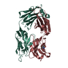
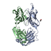
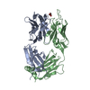
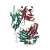
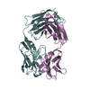
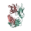
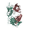
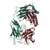
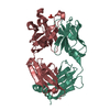
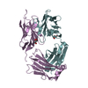
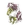
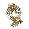
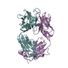
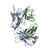
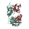
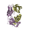
| Title | Crystal structure of humanized KR127 FAB | ||||||
 Components Components |
| ||||||
 Keywords Keywords | IMMUNE SYSTEM / HEPATITIS B VIRUS / HUMANIZED ANTIBODY / MONOCLONAL ANTIBODY / NEUTRALIZATION / PRES1 | ||||||
| Function / homology | Immunoglobulins / Immunoglobulin-like / Sandwich / Mainly Beta Function and homology information Function and homology information | ||||||
| Biological species |  | ||||||
| Method |  X-RAY DIFFRACTION / MOLECULAR REPLACEMENT / Resolution: 2.5 Å X-RAY DIFFRACTION / MOLECULAR REPLACEMENT / Resolution: 2.5 Å | ||||||
 Authors Authors | Chi, S.-W. / Kim, S.-J. / Maeng, C.-Y. / Hong, H.J. / Ryu, S.-E. | ||||||
 Citation Citation | Journal: Proc.Natl.Acad.Sci.Usa / Year: 2007 Title: Broadly neutralizing anti-hepatitis B virus antibody reveals a complementarity determining region H3 lid-opening mechanism Authors: Chi, S.-W. / Maeng, C.-Y. / Kim, S.J. / Oh, M.S. / Ryu, C.J. / Kim, S.-J. / Han, K.-H. / Hong, H.J. / Ryu, S.-E. | ||||||
| History |
|
- Structure visualization
| Structure viewer | Molecule: MolmilJmol/JSmol |
|---|
- Downloads & links
-Download
| PDBx/mmCIF format | 2eh7.cif.gz | 93.8 KB | Display | PDBx/mmCIF format |
|---|---|---|---|---|
| PDB format | pdb2eh7.ent.gz | 71.4 KB | Display | PDB format |
| PDBx/mmJSON format | 2eh7.json.gz | Tree view | PDBx/mmJSON format | |
| Others |  Other downloads Other downloads |
-Validation report
| Arichive directory | https://data.pdbj.org/pub/pdb/validation_reports/eh/2eh7ftp://data.pdbj.org/pub/pdb/validation_reports/eh/2eh7 | HTTPS FTP |
|---|
-Related structure data
| Related structure data |  2eh8C  1aj7S C: citing same article ( S: Starting model for refinement |
|---|---|
| Similar structure data |


-Links
 PDBj
PDBj

- Assembly
| Deposited unit | 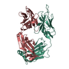
| ||||||||
|---|---|---|---|---|---|---|---|---|---|
| 1 |
| ||||||||
| Unit cell |
|
-Components
| #1: Antibody | Mass: 23891.732 Da / Num. of mol.: 1 Source method: isolated from a genetically manipulated source Source: (gene. exp.)  Homo sapiens (human) Homo sapiens (human) |
|---|---|
| #2: Antibody | Mass: 23086.756 Da / Num. of mol.: 1 Source method: isolated from a genetically manipulated source Source: (gene. exp.) Homo sapiens (human) |
| Has protein modification | Y |
| Sequence details | A SEQUENCE DATABASE REFERENCE FOR THIS PROTEIN DOES NOT CURRENTLY EXIST. THE SEQUENCES ARE SHOWN IN ...A SEQUENCE DATABASE REFERENCE FOR THIS PROTEIN DOES NOT CURRENTLY EXIST. THE SEQUENCES ARE SHOWN IN THE PAPER(FIGURE 1): HONG, H.J. ET AL., VIROLOGY 318, 134-141 (2004) |
-Experimental details
-Experiment
| Experiment | Method: X-RAY DIFFRACTION / Number of used crystals: 1 |
|---|
- Sample preparation
| Crystal | Density Matthews: 2.52 Å3/Da / Density % sol: 51.23 % |
|---|---|
| Crystal grow | Temperature: 288 K / Method: vapor diffusion, hanging drop / pH: 7.5 Details: 17% PEG 4000, 10mM Hepes-NaOH, 0.2M ammonium sulfate, pH 7.50, VAPOR DIFFUSION, HANGING DROP, temperature 288K |
-Data collection
| Diffraction | Mean temperature: 298 K |
|---|---|
| Diffraction source | Source: ROTATING ANODE / Type: RIGAKU RU300 / Wavelength: 1.5418 / Wavelength: 1.5418 Å |
| Detector | Type: RIGAKU RAXIS IV / Detector: IMAGE PLATE / Date: Jan 9, 2001 |
| Radiation | Monochromator: CONFOCAL MIRROR / Protocol: SINGLE WAVELENGTH / Monochromatic (M) / Laue (L): M / Scattering type: x-ray |
| Radiation wavelength | Wavelength: 1.5418 Å / Relative weight: 1 |
| Reflection | Resolution: 2.5→31.31 Å / Num. all: 16404 / Num. obs: 15758 / % possible obs: 96.3 % / Observed criterion σ(F): 0 / Observed criterion σ(I): 0 / Rmerge(I) obs: 0.078 / Net I/σ(I): 5.9 |
| Reflection shell | Resolution: 2.5→2.66 Å / % possible all: 97.5 |
- Processing
| Software |
| |||||||||||||||||||||||||
|---|---|---|---|---|---|---|---|---|---|---|---|---|---|---|---|---|---|---|---|---|---|---|---|---|---|---|
| Refinement | Method to determine structure: MOLECULAR REPLACEMENT Starting model: PDB ENTRY 1AJ7 Resolution: 2.5→31.31 Å / Cross valid method: THROUGHOUT / σ(F): 0 / σ(I): 0
| |||||||||||||||||||||||||
| Refinement step | Cycle: LAST / Resolution: 2.5→31.31 Å
| |||||||||||||||||||||||||
| Refine LS restraints |
| |||||||||||||||||||||||||
| LS refinement shell | Resolution: 2.5→2.66 Å / % reflection obs: 97.5 % |
