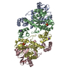
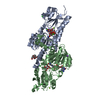
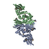
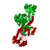
[English] 日本語
Yorodumi
- PDB-2b4j: Structural basis for the recognition between HIV-1 integrase and ... -

+
Open data


ID or keywords:

Loading...

-
Basic information

Entry
Database: PDB / ID: 2b4j
TitleStructural basis for the recognition between HIV-1 integrase and LEDGF/p75
Components
  • Integrase (IN)
  • PC4 and SFRS1 interacting protein
Keywordsviral protein / recombination / HIV / integration / transcription
Function / homology
Function and homology information


supercoiled DNA binding / Integration of viral DNA into host genomic DNA / Autointegration results in viral DNA circles / Formation of WDR5-containing histone-modifying complexes / 2-LTR circle formation / Vpr-mediated nuclear import of PICs / Integration of provirus / APOBEC3G mediated resistance to HIV-1 infection / mRNA 5'-splice site recognition / heterochromatin ...supercoiled DNA binding / Integration of viral DNA into host genomic DNA / Autointegration results in viral DNA circles / Formation of WDR5-containing histone-modifying complexes / 2-LTR circle formation / Vpr-mediated nuclear import of PICs / Integration of provirus / APOBEC3G mediated resistance to HIV-1 infection / mRNA 5'-splice site recognition / heterochromatin / nuclear periphery / euchromatin / HIV-1 retropepsin / : / retroviral ribonuclease H / exoribonuclease H / : / exoribonuclease H activity / host multivesicular body / DNA integration / RNA-directed DNA polymerase / viral genome integration into host DNA / viral penetration into host nucleus / establishment of integrated proviral latency / RNA-directed DNA polymerase activity / Transferases; Transferring phosphorus-containing groups; Nucleotidyltransferases / RNA-DNA hybrid ribonuclease activity / response to heat / viral nucleocapsid / DNA-binding transcription factor binding / DNA recombination / response to oxidative stress / Hydrolases; Acting on ester bonds / DNA-directed DNA polymerase / transcription coactivator activity / aspartic-type endopeptidase activity / DNA-directed DNA polymerase activity / chromatin remodeling / symbiont entry into host cell / symbiont-mediated suppression of host gene expression / lipid binding / host cell nucleus / chromatin binding / structural molecule activity / host cell plasma membrane / virion membrane / positive regulation of transcription by RNA polymerase II / proteolysis / DNA binding / RNA binding / zinc ion binding / nucleoplasm / membrane / nucleus / cytosol
Similarity search - Function
Conserved domain common to transcription factors TFIIS, elongin A, CRSP70 / Lens epithelium-derived growth factor, integrase-binding domain / HIV integrase-binding domain superfamily / Lens epithelium-derived growth factor (LEDGF) / Transcription Elongation Factor S-II; Chain A / TFIIS/LEDGF domain superfamily / domain with conserved PWWP motif / PWWP domain / PWWP domain profile. / PWWP domain ...Conserved domain common to transcription factors TFIIS, elongin A, CRSP70 / Lens epithelium-derived growth factor, integrase-binding domain / HIV integrase-binding domain superfamily / Lens epithelium-derived growth factor (LEDGF) / Transcription Elongation Factor S-II; Chain A / TFIIS/LEDGF domain superfamily / domain with conserved PWWP motif / PWWP domain / PWWP domain profile. / PWWP domain / Ribonuclease H-like superfamily/Ribonuclease H / Reverse transcriptase connection / Reverse transcriptase connection domain / Reverse transcriptase thumb / Reverse transcriptase thumb domain / Integrase Zinc binding domain / Zinc finger integrase-type profile. / Integrase-like, N-terminal / Integrase DNA binding domain / Integrase, C-terminal domain superfamily, retroviral / Integrase, N-terminal zinc-binding domain / Integrase, C-terminal, retroviral / Integrase DNA binding domain profile. / Immunodeficiency lentiviral matrix, N-terminal / gag gene protein p17 (matrix protein) / RNase H / Integrase core domain / Integrase, catalytic core / Integrase catalytic domain profile. / Retroviral nucleocapsid Gag protein p24, C-terminal domain / Gag protein p24 C-terminal domain / Retropepsin-like catalytic domain / Matrix protein, lentiviral and alpha-retroviral, N-terminal / Ribonuclease H domain / RNase H type-1 domain profile. / Reverse transcriptase (RNA-dependent DNA polymerase) / Reverse transcriptase domain / Reverse transcriptase (RT) catalytic domain profile. / Retropepsins / Retroviral aspartyl protease / Aspartyl protease, retroviral-type family profile. / Peptidase A2A, retrovirus, catalytic / Retrovirus capsid, C-terminal / Retroviral matrix protein / Retrovirus capsid, N-terminal / zinc finger / Zinc knuckle / Zinc finger, CCHC-type superfamily / Zinc finger, CCHC-type / Zinc finger CCHC-type profile. / Nucleotidyltransferase; domain 5 / Ribonuclease H superfamily / Aspartic peptidase, active site / Eukaryotic and viral aspartyl proteases active site. / Aspartic peptidase domain superfamily / Ribonuclease H-like superfamily / Reverse transcriptase/Diguanylate cyclase domain / DNA/RNA polymerase superfamily / Up-down Bundle / 2-Layer Sandwich / Mainly Alpha / Alpha Beta
Similarity search - Domain/homology
PHOSPHATE ION / PC4 and SFRS1-interacting protein / Gag-Pol polyprotein
Similarity search - Component
Biological speciesHuman immunodeficiency virus 1
Homo sapiens (human)
MethodX-RAY DIFFRACTION / SYNCHROTRON / MOLECULAR REPLACEMENT / Resolution: 2.02 Å
AuthorsCherepanov, P. / Ambrosio, A.L. / Rahman, S. / Ellenberger, T. / Engelman, A.
CitationJournal: Proc.Natl.Acad.Sci.Usa / Year: 2005
Title: Structural basis for the recognition between HIV-1 integrase and transcriptional coactivator p75
Authors: Cherepanov, P. / Ambrosio, A.L. / Rahman, S. / Ellenberger, T. / Engelman, A.
History
DepositionSep 24, 2005Deposition site: RCSB / Processing site: RCSB
Revision 1.0Oct 25, 2005Provider: repository / Type: Initial release
Revision 1.1May 1, 2008Group: Version format compliance
Revision 1.2Jul 13, 2011Group: Advisory / Version format compliance
Revision 1.3Oct 11, 2017Group: Advisory / Refinement description / Category: pdbx_unobs_or_zero_occ_atoms / software
Item: _software.classification / _software.contact_author ..._software.classification / _software.contact_author / _software.contact_author_email / _software.date / _software.language / _software.location / _software.name / _software.type / _software.version
Revision 1.4Oct 20, 2021Group: Advisory / Database references / Derived calculations
Category: database_2 / pdbx_unobs_or_zero_occ_atoms ...database_2 / pdbx_unobs_or_zero_occ_atoms / struct_ref_seq_dif / struct_site
Item: _database_2.pdbx_DOI / _database_2.pdbx_database_accession ..._database_2.pdbx_DOI / _database_2.pdbx_database_accession / _struct_ref_seq_dif.details / _struct_site.pdbx_auth_asym_id / _struct_site.pdbx_auth_comp_id / _struct_site.pdbx_auth_seq_id
Revision 1.5Aug 23, 2023Group: Data collection / Refinement description
Category: chem_comp_atom / chem_comp_bond / pdbx_initial_refinement_model

-
Structure visualization

Structure viewerMolecule:
MolmilJmol/JSmol

Downloads & links

-
Assembly

Deposited unit
A: Integrase (IN)
B: Integrase (IN)
C: PC4 and SFRS1 interacting protein
D: PC4 and SFRS1 interacting protein
hetero molecules


Theoretical massNumber of molelcules
Total (without water)59,52410
Polymers58,9664
Non-polymers5586
Water3,963220
1


  • Idetical with deposited unit
  • defined by author&software
TypeNameSymmetry operationNumber
identity operation1_555x,y,z1
Buried area6690 Å2
ΔGint-41 kcal/mol
Surface area20310 Å2
MethodPISA
2
A: Integrase (IN)
B: Integrase (IN)
C: PC4 and SFRS1 interacting protein
D: PC4 and SFRS1 interacting protein
hetero molecules

A: Integrase (IN)
B: Integrase (IN)
C: PC4 and SFRS1 interacting protein
D: PC4 and SFRS1 interacting protein
hetero molecules


Theoretical massNumber of molelcules
Total (without water)119,04820
Polymers117,9328
Non-polymers1,11712
Water1448
TypeNameSymmetry operationNumber
identity operation1_555x,y,z1
crystal symmetry operation2_555-x,y,-z1
Buried area13640 Å2
ΔGint-78 kcal/mol
Surface area40360 Å2
MethodPISA
Unit cell
Length a, b, c (Å)122.421, 60.593, 71.126
Angle α, β, γ (deg.)90.00, 109.06, 90.00
Int Tables number5
Space group name H-MC121

-
Components

#1: Protein Integrase (IN)


Mass: 18194.760 Da / Num. of mol.: 2 / Fragment: HIV-1 integrase / Mutation: F185K
Source method: isolated from a genetically manipulated source
Source: (gene. exp.) Human immunodeficiency virus 1 / Genus: Lentivirus / Gene: POL / Plasmid: pET15b / Species (production host): Escherichia coli / Production host: Escherichia coli BL21(DE3) (bacteria) / Strain (production host): BL21(DE3) / References: UniProt: P12497
#2: Protein PC4 and SFRS1 interacting protein / Lens epithelium-derived growth factor / Transcriptional coactivator p75/p52 / Dense fine speckles ...Lens epithelium-derived growth factor / Transcriptional coactivator p75/p52 / Dense fine speckles 70 kDa protein / DFS 70 / CLL-associated antigen KW-7


Mass: 11288.199 Da / Num. of mol.: 2 / Fragment: LEDGF
Source method: isolated from a genetically manipulated source
Source: (gene. exp.) Homo sapiens (human) / Gene: PSIP1, LEDGF / Plasmid: pCPGST75-81 / Species (production host): Escherichia coli / Production host: Escherichia coli BL21 (bacteria) / Strain (production host): BL21 / References: UniProt: O75475
#3: Chemical ChemComp-PO4 / PHOSPHATE ION / Phosphate


Mass: 94.971 Da / Num. of mol.: 2 / Source method: obtained synthetically / Formula: PO4
#4: Chemical
ChemComp-GOL / GLYCEROL / GLYCERIN / PROPANE-1,2,3-TRIOL / Glycerol


Mass: 92.094 Da / Num. of mol.: 4 / Source method: obtained synthetically / Formula: C3H8O3
#5: Water ChemComp-HOH / water / Water


Mass: 18.015 Da / Num. of mol.: 220 / Source method: isolated from a natural source / Formula: H2O

-
Experimental details

-
Experiment

ExperimentMethod: X-RAY DIFFRACTION / Number of used crystals: 1

-
Sample preparation

CrystalDensity Matthews: 2.12 Å3/Da / Density % sol: 41.87 %
Crystal growTemperature: 295 K / Method: vapor diffusion, hanging drop / pH: 6.25
Details: PEG-3350, Na2HPO4, NaH2PO4, KH2PO4, NACL, HEPES, pH 6.25, VAPOR DIFFUSION, HANGING DROP, temperature 295K

-
Data collection

DiffractionMean temperature: 100 K
Diffraction sourceSource: SYNCHROTRON / Site: NSLS / Beamline: X26C / Wavelength: 1 Å
DetectorType: ADSC QUANTUM 4 / Detector: CCD / Date: Mar 26, 2005
RadiationProtocol: SINGLE WAVELENGTH / Monochromatic (M) / Laue (L): M / Scattering type: x-ray
Radiation wavelengthWavelength: 1 Å / Relative weight: 1
ReflectionResolution: 2.02→50 Å / Num. all: 32648 / Num. obs: 32136 / % possible obs: 99.2 % / Observed criterion σ(F): 3.1 / Observed criterion σ(I): 3.1 / Redundancy: 4.8 % / Biso Wilson estimate: 35.2 Å2 / Rmerge(I) obs: 0.072 / Χ2: 1.041 / Net I/σ(I): 18.9
Reflection shellResolution: 2.02→2.09 Å / % possible obs: 99.2 % / Redundancy: 4.5 % / Rmerge(I) obs: 0.47 / Mean I/σ(I) obs: 3.1 / Num. measured obs: 3189 / Χ2: 1.007 / % possible all: 99.2

-
Processing

Software
NameVersionClassificationNB
DENZOdata reduction
SCALEPACKdata scaling
REFMACrefinement
PDB_EXTRACT1.7data extraction
CBASSdata collection
CCP4phasing
RefinementMethod to determine structure: MOLECULAR REPLACEMENT
Starting model: PDB Entry: 1BIS (Integrase), 1Z9E (LEDGF)
Resolution: 2.02→20 Å / Cor.coef. Fo:Fc: 0.957 / Cor.coef. Fo:Fc free: 0.942 / SU B: 8.106 / SU ML: 0.115 / TLS residual ADP flag: LIKELY RESIDUAL / Cross valid method: THROUGHOUT / σ(F): 0 / ESU R: 0.184 / ESU R Free: 0.164 / Stereochemistry target values: MAXIMUM LIKELIHOOD
RfactorNum. reflection% reflectionSelection details
Rfree0.226 1627 5.1 %RANDOM
Rwork0.181 ---
all0.183 30406 --
obs0.183 32113 99.19 %-
Solvent computationIon probe radii: 0.8 Å / Shrinkage radii: 0.8 Å / VDW probe radii: 1.2 Å / Solvent model: MASK
Displacement parametersBiso mean: 37.386 Å2
Baniso -1Baniso -2Baniso -3
1-0.03 Å20 Å21.3 Å2
2---0.38 Å20 Å2
3---1.2 Å2
Refinement stepCycle: LAST / Resolution: 2.02→20 Å
ProteinNucleic acidLigandSolventTotal
Num. atoms3503 0 34 220 3757
Refine LS restraints
Refine-IDTypeDev idealDev ideal targetNumber
X-RAY DIFFRACTIONr_bond_refined_d0.0170.0223584
X-RAY DIFFRACTIONr_angle_refined_deg1.6021.9524819
X-RAY DIFFRACTIONr_dihedral_angle_1_deg5.5245437
X-RAY DIFFRACTIONr_dihedral_angle_2_deg32.6925.033151
X-RAY DIFFRACTIONr_dihedral_angle_3_deg15.92215693
X-RAY DIFFRACTIONr_dihedral_angle_4_deg19.5381517
X-RAY DIFFRACTIONr_chiral_restr0.1320.2559
X-RAY DIFFRACTIONr_gen_planes_refined0.0070.022553
X-RAY DIFFRACTIONr_nbd_refined0.2130.21687
X-RAY DIFFRACTIONr_nbtor_refined0.3010.22521
X-RAY DIFFRACTIONr_xyhbond_nbd_refined0.1470.2222
X-RAY DIFFRACTIONr_symmetry_vdw_refined0.1790.232
X-RAY DIFFRACTIONr_symmetry_hbond_refined0.2370.212
X-RAY DIFFRACTIONr_mcbond_it0.9811.52295
X-RAY DIFFRACTIONr_mcangle_it1.623564
X-RAY DIFFRACTIONr_scbond_it2.77831487
X-RAY DIFFRACTIONr_scangle_it4.3614.51255
LS refinement shellResolution: 2.021→2.073 Å / Total num. of bins used: 20
RfactorNum. reflection% reflection
Rfree0.276 124 -
Rwork0.214 2223 -
all-2347 -
obs--98.32 %
Refinement TLS params.

Method: refined / Refine-ID: X-RAY DIFFRACTION

IDL112)L122)L132)L222)L232)L332)S11 (Å °)S12 (Å °)S13 (Å °)S21 (Å °)S22 (Å °)S23 (Å °)S31 (Å °)S32 (Å °)S33 (Å °)T112)T122)T132)T222)T232)T332)Origin x (Å)Origin y (Å)Origin z (Å)
14.4292-0.5959-1.02571.74120.39171.82380.21540.34570.5314-0.0421-0.0868-0.0325-0.1932-0.0167-0.1286-0.06550.01260.0322-0.07160.0744-0.04690.8516-0.4794-21.0989
24.1647-0.3954-1.30460.9899-0.07161.6376-0.07120.3343-0.39680.0429-0.03660.23310.0937-0.25250.1078-0.0695-0.01030.0048-0.0585-0.0501-0.0106-13.536-12.9638-15.861
32.28330.56980.36085.12921.56672.5546-0.21460.16980.011-0.40250.17930.2188-0.0725-0.04910.0353-0.1407-0.0269-0.0106-0.1154-0.0025-0.1097-25.320519.5794-4.2029
42.6236-1.4640.5096.3798-3.1633.92440.13390.3782-0.1065-0.9928-0.126-0.04260.47440.0114-0.0079-0.03280.0122-0.0248-0.02830.0133-0.141618.1455-31.5844-18.216
Refinement TLS group

Refine-ID: X-RAY DIFFRACTION / Selection: ALL

IDRefine TLS-IDAuth asym-IDLabel asym-IDAuth seq-IDLabel seq-ID
11AA56 - 20810 - 162
22BB57 - 20811 - 162
33CC345 - 4261 - 82
44DD345 - 4261 - 82

+
About Yorodumi

-
News

-
Feb 9, 2022. New format data for meta-information of EMDB entries

New format data for meta-information of EMDB entries

  • Version 3 of the EMDB header file is now the official format.
  • The previous official version 1.9 will be removed from the archive.

Related info.:EMDB header

External links:wwPDB to switch to version 3 of the EMDB data model

-
Aug 12, 2020. Covid-19 info

Covid-19 info

URL: https://pdbj.org/emnavi/covid19.php

New page: Covid-19 featured information page in EM Navigator.

Related info.:Covid-19 info / Mar 5, 2020. Novel coronavirus structure data

+
Mar 5, 2020. Novel coronavirus structure data

Novel coronavirus structure data

Related info.:Yorodumi Speices / Aug 12, 2020. Covid-19 info

External links:COVID-19 featured content - PDBj / Molecule of the Month (242):Coronavirus Proteases

+
Jan 31, 2019. EMDB accession codes are about to change! (news from PDBe EMDB page)

EMDB accession codes are about to change! (news from PDBe EMDB page)

  • The allocation of 4 digits for EMDB accession codes will soon come to an end. Whilst these codes will remain in use, new EMDB accession codes will include an additional digit and will expand incrementally as the available range of codes is exhausted. The current 4-digit format prefixed with “EMD-” (i.e. EMD-XXXX) will advance to a 5-digit format (i.e. EMD-XXXXX), and so on. It is currently estimated that the 4-digit codes will be depleted around Spring 2019, at which point the 5-digit format will come into force.
  • The EM Navigator/Yorodumi systems omit the EMD- prefix.

Related info.:Q: What is EMD? / ID/Accession-code notation in Yorodumi/EM Navigator

External links:EMDB Accession Codes are Changing Soon! / Contact to PDBj

+
Jul 12, 2017. Major update of PDB

Major update of PDB

  • wwPDB released updated PDB data conforming to the new PDBx/mmCIF dictionary.
  • This is a major update changing the version number from 4 to 5, and with Remediation, in which all the entries are updated.
  • In this update, many items about electron microscopy experimental information are reorganized (e.g. em_software).
  • Now, EM Navigator and Yorodumi are based on the updated data.

External links:wwPDB Remediation / Enriched Model Files Conforming to OneDep Data Standards Now Available in the PDB FTP Archive

-
Yorodumi

Thousand views of thousand structures

  • Yorodumi is a browser for structure data from EMDB, PDB, SASBDB, etc.
  • This page is also the successor to EM Navigator detail page, and also detail information page/front-end page for Omokage search.
  • The word "yorodu" (or yorozu) is an old Japanese word meaning "ten thousand". "mi" (miru) is to see.

Related info.:EMDB / PDB / SASBDB / Comparison of 3 databanks / Yorodumi Search / Aug 31, 2016. New EM Navigator & Yorodumi / Yorodumi Papers / Jmol/JSmol / Function and homology information / Changes in new EM Navigator and Yorodumi

Read more