 Movie
Movie Controller

[English] 日本語
 Yorodumi
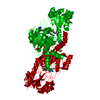
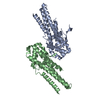
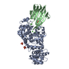
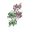
- PDB-5tbf: Crystal structure of SeMet derivatives of domain2 and domain 3 of RctB -
+ Open data

- Basic information
| Entry | Database: PDB / ID: 5tbf | ||||||
|---|---|---|---|---|---|---|---|
| Title | Crystal structure of SeMet derivatives of domain2 and domain 3 of RctB | ||||||
 Components Components | Translation elongation factor | ||||||
 Keywords Keywords | TRANSLATION / RctB | ||||||
| Function / homology | Replication initiator protein RctB, central region / RctB, helix turn helix domain / Vibrionales, replication initiator protein RctB, central region / RctB helix turn helix domain / translation elongation factor activity / Translation elongation factor / Uncharacterized protein Function and homology information Function and homology information | ||||||
| Biological species |   Vibrio cholerae (bacteria) Vibrio cholerae (bacteria) | ||||||
| Method |  X-RAY DIFFRACTION / SYNCHROTRON / SAD / Resolution: 3.003 Å X-RAY DIFFRACTION / SYNCHROTRON / SAD / Resolution: 3.003 Å | ||||||
 Authors Authors | Li, M. / Wlodawer, A. / Chattoraj, D. / Jha, J. | ||||||
 Citation Citation | Journal: To Be Published Title: Crystal Structure of core domains of RctB Authors: Li, M. / Wlodawer, A. / Jha, J. / Chattoraj, D. / Gustchina, A. | ||||||
| History |
|
- Structure visualization
| Structure viewer | Molecule: MolmilJmol/JSmol |
|---|
- Downloads & links
-Download
| PDBx/mmCIF format | 5tbf.cif.gz | 124.8 KB | Display | PDBx/mmCIF format |
|---|---|---|---|---|
| PDB format | pdb5tbf.ent.gz | 98.1 KB | Display | PDB format |
| PDBx/mmJSON format | 5tbf.json.gz | Tree view | PDBx/mmJSON format | |
| Others |  Other downloads Other downloads |
-Validation report
| Arichive directory | https://data.pdbj.org/pub/pdb/validation_reports/tb/5tbfftp://data.pdbj.org/pub/pdb/validation_reports/tb/5tbf | HTTPS FTP |
|---|
-Related structure data
| Similar structure data |
|---|


-Links
 PDBj
PDBj- Assembly
| Deposited unit | 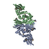
| ||||||||
|---|---|---|---|---|---|---|---|---|---|
| 1 |
| ||||||||
| Unit cell |
|
-Components
| #1: Protein | Mass: 38469.270 Da / Num. of mol.: 2 / Fragment: residues 145-470 Source method: isolated from a genetically manipulated source Source: (gene. exp.) Vibrio cholerae (bacteria) / Gene: ERS013166_00020 / Plasmid: pJJ399 / Cell (production host): Bl21De3 / Production host: Has protein modification | Y | |
|---|
-Experimental details
-Experiment
| Experiment | Method: X-RAY DIFFRACTION / Number of used crystals: 1 |
|---|
- Sample preparation
| Crystal | Density Matthews: 2.91 Å3/Da / Density % sol: 57.73 % |
|---|---|
| Crystal grow | Method: vapor diffusion, hanging drop / pH: 4.5 / Details: 1.5M Ammonium Sulfate |
-Data collection
| Diffraction | Mean temperature: 100 K |
|---|---|
| Diffraction source | Source: SYNCHROTRON / Site: APS  / Beamline: 22-ID / Wavelength: 0.9786 Å / Beamline: 22-ID / Wavelength: 0.9786 Å |
| Detector | Type: RAYONIX MX300-HS / Detector: CCD / Date: Aug 19, 2016 |
| Radiation | Protocol: SINGLE WAVELENGTH / Monochromatic (M) / Laue (L): M / Scattering type: x-ray |
| Radiation wavelength | Wavelength: 0.9786 Å / Relative weight: 1 |
| Reflection | Resolution: 3→55 Å / Num. obs: 18054 / % possible obs: 99.1 % / Redundancy: 6.7 % / Net I/σ(I): 19.68 |
- Processing
| Software |
| |||||||||||||||||||||||||||||||||||||||||||||||||
|---|---|---|---|---|---|---|---|---|---|---|---|---|---|---|---|---|---|---|---|---|---|---|---|---|---|---|---|---|---|---|---|---|---|---|---|---|---|---|---|---|---|---|---|---|---|---|---|---|---|---|
| Refinement | Method to determine structure: SAD / Resolution: 3.003→43.37 Å / SU ML: 0.41 / Cross valid method: NONE / σ(F): 1.35 / Phase error: 28.06 / Stereochemistry target values: MLHL
| |||||||||||||||||||||||||||||||||||||||||||||||||
| Solvent computation | Shrinkage radii: 0.9 Å / VDW probe radii: 1.11 Å / Solvent model: FLAT BULK SOLVENT MODEL | |||||||||||||||||||||||||||||||||||||||||||||||||
| Refinement step | Cycle: LAST / Resolution: 3.003→43.37 Å
| |||||||||||||||||||||||||||||||||||||||||||||||||
| Refine LS restraints |
| |||||||||||||||||||||||||||||||||||||||||||||||||
| LS refinement shell |
|
