 Movie
Movie Controller

[English] 日本語
 Yorodumi
- PDB-2as5: Structure of the DNA binding domains of NFAT and FOXP2 bound spec... -
+ Open data

- Basic information
| Entry | Database: PDB / ID: 2as5 | ||||||
|---|---|---|---|---|---|---|---|
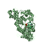
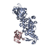
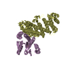
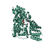
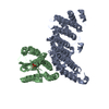
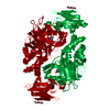
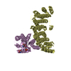
| Title | Structure of the DNA binding domains of NFAT and FOXP2 bound specifically to DNA. | ||||||
 Components Components |
| ||||||
 Keywords Keywords | Transcription/DNA / Forkhead Domain / RHR Domain / Rel Homology Region / IG Fold / Winged Helix-Turn-Helix / B-DNA / Transcription-DNA COMPLEX | ||||||
| Function / homology |  Function and homology information Function and homology informationcaudate nucleus development / putamen development / transcription factor AP-1 complex / negative regulation of vascular associated smooth muscle cell differentiation / myotube cell development / RUNX1 and FOXP3 control the development of regulatory T lymphocytes (Tregs) / calcineurin-NFAT signaling cascade / cartilage development / CLEC7A (Dectin-1) induces NFAT activation / positive regulation of myoblast fusion ...caudate nucleus development / putamen development / transcription factor AP-1 complex / negative regulation of vascular associated smooth muscle cell differentiation / myotube cell development / RUNX1 and FOXP3 control the development of regulatory T lymphocytes (Tregs) / calcineurin-NFAT signaling cascade / cartilage development / CLEC7A (Dectin-1) induces NFAT activation / positive regulation of myoblast fusion / Calcineurin activates NFAT / phosphatase binding / positive regulation of B cell proliferation / 14-3-3 protein binding / cellular response to calcium ion / FCERI mediated Ca+2 mobilization / B cell receptor signaling pathway / DNA-binding transcription repressor activity, RNA polymerase II-specific / cerebral cortex development / sequence-specific double-stranded DNA binding / cell migration / DNA-binding transcription activator activity, RNA polymerase II-specific / transcription regulator complex / sequence-specific DNA binding / transcription by RNA polymerase II / molecular adaptor activity / DNA-binding transcription factor activity, RNA polymerase II-specific / ribonucleoprotein complex / response to xenobiotic stimulus / RNA polymerase II cis-regulatory region sequence-specific DNA binding / DNA-binding transcription factor activity / negative regulation of DNA-templated transcription / chromatin binding / DNA damage response / regulation of transcription by RNA polymerase II / positive regulation of gene expression / positive regulation of DNA-templated transcription / chromatin / regulation of DNA-templated transcription / protein homodimerization activity / DNA binding / nucleoplasm / identical protein binding / nucleus / metal ion binding / cytoplasm / cytosol Similarity search - Function | ||||||
| Biological species |  Homo sapiens (human) Homo sapiens (human) | ||||||
| Method |  X-RAY DIFFRACTION / SYNCHROTRON / MOLECULAR REPLACEMENT / Resolution: 2.7 Å X-RAY DIFFRACTION / SYNCHROTRON / MOLECULAR REPLACEMENT / Resolution: 2.7 Å | ||||||
 Authors Authors | Wu, Y. / Stroud, J.C. / Borde, M. / Bates, D.L. / Guo, L. / Han, A. / Rao, A. / Chen, L. | ||||||
 Citation Citation | Journal: Cell(Cambridge,Mass.) / Year: 2006 Title: FOXP3 Controls Regulatory T Cell Function through Cooperation with NFAT. Authors: Wu, Y. / Borde, M. / Heissmeyer, V. / Feuerer, M. / Lapan, A.D. / Stroud, J.C. / Bates, D.L. / Guo, L. / Han, A. / Ziegler, S.F. / Mathis, D. / Benoist, C. / Chen, L. / Rao, A. | ||||||
| History |
|
- Structure visualization
| Structure viewer | Molecule: MolmilJmol/JSmol |
|---|
- Downloads & links
-Download
| PDBx/mmCIF format | 2as5.cif.gz | 211.4 KB | Display | PDBx/mmCIF format |
|---|---|---|---|---|
| PDB format | pdb2as5.ent.gz | 162.9 KB | Display | PDB format |
| PDBx/mmJSON format | 2as5.json.gz | Tree view | PDBx/mmJSON format | |
| Others |  Other downloads Other downloads |
-Validation report
| Summary document | 2as5_validation.pdf.gz | 484.3 KB | Display | wwPDB validaton report |
|---|---|---|---|---|
| Full document | 2as5_full_validation.pdf.gz | 526.3 KB | Display | |
| Data in XML | 2as5_validation.xml.gz | 35.7 KB | Display | |
| Data in CIF | 2as5_validation.cif.gz | 49.3 KB | Display | |
| Arichive directory | https://data.pdbj.org/pub/pdb/validation_reports/as/2as5ftp://data.pdbj.org/pub/pdb/validation_reports/as/2as5 | HTTPS FTP |
-Related structure data
| Related structure data |  1a02S S: Starting model for refinement |
|---|---|
| Similar structure data |


-Links
 PDBj
PDBj


- Assembly
| Deposited unit | 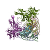
| ||||||||
|---|---|---|---|---|---|---|---|---|---|
| 1 | 
| ||||||||
| 2 | 
| ||||||||
| Unit cell |
|
-Components
-DNA chain , 2 types, 4 molecules ACBD
| #1: DNA chain | Mass: 6491.230 Da / Num. of mol.: 2 / Source method: obtained synthetically / Details: Solid phase synthesis #2: DNA chain | Mass: 6389.186 Da / Num. of mol.: 2 / Source method: obtained synthetically |
|---|
-Protein , 2 types, 4 molecules NMFG
| #3: Protein | Mass: 32582.117 Da / Num. of mol.: 2 / Fragment: NFAT1 DNA BINDING DOMAIN Source method: isolated from a genetically manipulated source Source: (gene. exp.) Homo sapiens (human) / Gene: NFATC2, NFAT1, NFATP / Plasmid: pLM1 / Species (production host): Escherichia coli / Production host:  #4: Protein | Mass: 11025.630 Da / Num. of mol.: 2 / Fragment: FOXP2 DNA BINDING DOMAIN / Mutation: D502I Source method: isolated from a genetically manipulated source Source: (gene. exp.) Homo sapiens (human) / Gene: FOXP2 / Plasmid: pET-30 LIC / Production host: |
|---|
-Non-polymers , 2 types, 117 molecules 

| #5: Chemical |  Mass: 24.305 Da / Num. of mol.: 2 / Source method: obtained synthetically / Formula: Mg Mass: 24.305 Da / Num. of mol.: 2 / Source method: obtained synthetically / Formula: Mg#6: Water | ChemComp-HOH / | Mass: 18.015 Da / Num. of mol.: 115 / Source method: isolated from a natural source / Formula: H2O |
|---|
-Experimental details
-Experiment
| Experiment | Method: X-RAY DIFFRACTION / Number of used crystals: 1 |
|---|
- Sample preparation
| Crystal | Density Matthews: 2.9 Å3/Da / Density % sol: 57.58 % | ||||||||||||||||||||||||||||||||||||||||||||
|---|---|---|---|---|---|---|---|---|---|---|---|---|---|---|---|---|---|---|---|---|---|---|---|---|---|---|---|---|---|---|---|---|---|---|---|---|---|---|---|---|---|---|---|---|---|
| Crystal grow | Temperature: 291 K / Method: vapor diffusion, hanging drop / pH: 6.3 Details: Cacodylic Acid, PEG 4k, Sodium Chloride, Magnesium Chloride, Glycerol, pH 6.3, VAPOR DIFFUSION, HANGING DROP, temperature 291K | ||||||||||||||||||||||||||||||||||||||||||||
| Components of the solutions |
|
-Data collection
| Diffraction | Mean temperature: 100 K |
|---|---|
| Diffraction source | Source: SYNCHROTRON / Site: ALS  / Beamline: 8.2.1 / Wavelength: 1.107 Å / Beamline: 8.2.1 / Wavelength: 1.107 Å |
| Detector | Type: ADSC QUANTUM 315 / Detector: CCD / Date: Dec 22, 2003 |
| Radiation | Monochromator: Double crystal, Si(111) / Protocol: SINGLE WAVELENGTH / Monochromatic (M) / Laue (L): M / Scattering type: x-ray |
| Radiation wavelength | Wavelength: 1.107 Å / Relative weight: 1 |
| Reflection | Resolution: 2.7→30 Å / Num. all: 32927 / Num. obs: 31620 / % possible obs: 96 % / Observed criterion σ(F): 0 / Observed criterion σ(I): 0 / Rmerge(I) obs: 0.085 / Net I/σ(I): 10.6 |
| Reflection shell | Resolution: 2.7→2.8 Å / % possible all: 86.5 |
- Processing
| Software |
| ||||||||||||||||||||
|---|---|---|---|---|---|---|---|---|---|---|---|---|---|---|---|---|---|---|---|---|---|
| Refinement | Method to determine structure: MOLECULAR REPLACEMENT Starting model: pdb entry 1a02, chain N Resolution: 2.7→30 Å / σ(F): 2 / Stereochemistry target values: Engh & Huber Details: Data was collected to 39.3 Angstroms. Resolutions lower than 30 was not included for refinement
| ||||||||||||||||||||
| Refine analyze | Luzzati coordinate error obs: 0.45 Å / Luzzati sigma a obs: 0.6 Å | ||||||||||||||||||||
| Refinement step | Cycle: LAST / Resolution: 2.7→30 Å
| ||||||||||||||||||||
| Refine LS restraints |
|
