 Movie
Movie Controller

[English] 日本語
 Yorodumi
- PDB-1ysa: THE GCN4 BASIC REGION LEUCINE ZIPPER BINDS DNA AS A DIMER OF UNIN... -
+ Open data

- Basic information
| Entry | Database: PDB / ID: 1ysa | ||||||
|---|---|---|---|---|---|---|---|
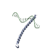
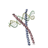
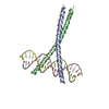
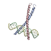
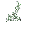
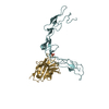
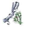
| Title | THE GCN4 BASIC REGION LEUCINE ZIPPER BINDS DNA AS A DIMER OF UNINTERRUPTED ALPHA HELICES: CRYSTAL STRUCTURE OF THE PROTEIN-DNA COMPLEX | ||||||
 Components Components |
| ||||||
 Keywords Keywords | TRANSCRIPTION/DNA /  PROTEIN-DNA COMPLEX / DOUBLE HELIX / TRANSCRIPTION-DNA COMPLEX PROTEIN-DNA COMPLEX / DOUBLE HELIX / TRANSCRIPTION-DNA COMPLEX | ||||||
| Function / homology |  Function and homology information Function and homology informationprotein localization to nuclear periphery / Activation of the AP-1 family of transcription factors / response to amino acid starvation / mediator complex binding / negative regulation of ribosomal protein gene transcription by RNA polymerase II / positive regulation of cellular response to amino acid starvation / nitrogen catabolite activation of transcription from RNA polymerase II promoter / TFIID-class transcription factor complex binding / amino acid biosynthetic process / positive regulation of transcription initiation by RNA polymerase II ...protein localization to nuclear periphery / Activation of the AP-1 family of transcription factors / response to amino acid starvation / mediator complex binding / negative regulation of ribosomal protein gene transcription by RNA polymerase II / positive regulation of cellular response to amino acid starvation / nitrogen catabolite activation of transcription from RNA polymerase II promoter / TFIID-class transcription factor complex binding / amino acid biosynthetic process / positive regulation of transcription initiation by RNA polymerase II / positive regulation of RNA polymerase II transcription preinitiation complex assembly / cellular response to amino acid starvation / RNA polymerase II transcription regulator complex / : / DNA-binding transcription activator activity, RNA polymerase II-specific / transcription regulator complex / RNA polymerase II-specific DNA-binding transcription factor binding / sequence-specific DNA binding / DNA-binding transcription factor activity, RNA polymerase II-specific / intracellular signal transduction / RNA polymerase II cis-regulatory region sequence-specific DNA binding / DNA-binding transcription factor activity / chromatin binding / negative regulation of transcription by RNA polymerase II / positive regulation of transcription by RNA polymerase II / identical protein binding / nucleusSimilarity search - Function | ||||||
| Biological species |  Saccharomyces cerevisiae (brewer's yeast) Saccharomyces cerevisiae (brewer's yeast) | ||||||
| Method | X-RAY DIFFRACTION / Resolution: 2.9 Å | ||||||
 Authors Authors | Ellenberger, T.E. / Brandl, C.J. / Struhl, K. / Harrison, S.C. | ||||||
 Citation Citation | Journal: Cell(Cambridge,Mass.) / Year: 1992 Title: The GCN4 basic region leucine zipper binds DNA as a dimer of uninterrupted alpha helices: crystal structure of the protein-DNA complex. Authors: Ellenberger, T.E. / Brandl, C.J. / Struhl, K. / Harrison, S.C. #1: Journal: Science / Year: 1991Title: X-Ray Structure of the GCN4 Leucine Zipper, a Two-Stranded, Parallel Coiled Coil Authors: O'Shea, E.K. / Klemm, J.D. / Kim, P.S. / Alber, T. | ||||||
| History |
|
- Structure visualization
| Structure viewer | Molecule: MolmilJmol/JSmol |
|---|
- Downloads & links
-Download
| PDBx/mmCIF format | 1ysa.cif.gz | 60.6 KB | Display | PDBx/mmCIF format |
|---|---|---|---|---|
| PDB format | pdb1ysa.ent.gz | 40.2 KB | Display | PDB format |
| PDBx/mmJSON format | 1ysa.json.gz | Tree view | PDBx/mmJSON format | |
| Others |  Other downloads Other downloads |
-Validation report
| Arichive directory | https://data.pdbj.org/pub/pdb/validation_reports/ys/1ysaftp://data.pdbj.org/pub/pdb/validation_reports/ys/1ysa | HTTPS FTP |
|---|
-Related structure data
| Similar structure data |
|---|

-Links
 PDBj
PDBj


- Assembly
| Deposited unit | 
| ||||||||
|---|---|---|---|---|---|---|---|---|---|
| 1 |
| ||||||||
| Unit cell |
| ||||||||
| Atom site foot note | 1: TWO ADENINES, B 22 AND B 23, ARE NEAR THE 5-PRIME END OF DNA CHAIN B AND ARE LOOPED OUT OF THE DNA HELIX. THIS CONFORMATION IS PRESENT IN CRYSTALS THAT WERE SOAKED IN 35% (VOL/VOL) POLYETHYLENE ...1: TWO ADENINES, B 22 AND B 23, ARE NEAR THE 5-PRIME END OF DNA CHAIN B AND ARE LOOPED OUT OF THE DNA HELIX. THIS CONFORMATION IS PRESENT IN CRYSTALS THAT WERE SOAKED IN 35% (VOL/VOL) POLYETHYLENE GLYCOL MW 400, FOLLOWED BY RAPID FREEZING AND X-RAY DATA COLLECTION AT -155 DEGREES CENTIGRADE. |
-Components
| #1: DNA chain | Mass: 6034.915 Da / Num. of mol.: 1 / Source method: obtained synthetically | ||
|---|---|---|---|
| #2: DNA chain | Mass: 6231.067 Da / Num. of mol.: 1 / Source method: obtained synthetically | ||
| #3: Protein | Mass: 6892.072 Da / Num. of mol.: 2 / Source method: isolated from a natural source / Source: (natural) Saccharomyces cerevisiae (brewer's yeast) / References: UniProt: P03069#4: Water | ChemComp-HOH / | Water Mass: 18.015 Da / Num. of mol.: 17 / Source method: isolated from a natural source / Formula: H2O Mass: 18.015 Da / Num. of mol.: 17 / Source method: isolated from a natural source / Formula: H2O |
-Experimental details
-Experiment
| Experiment | Method: X-RAY DIFFRACTION |
|---|
- Sample preparation
| Crystal | Density Matthews: 2.43 Å3/Da / Density % sol: 49.4 % | ||||||||||||||||||||||||||||||||||||||||||||||||||||||||||||||||||
|---|---|---|---|---|---|---|---|---|---|---|---|---|---|---|---|---|---|---|---|---|---|---|---|---|---|---|---|---|---|---|---|---|---|---|---|---|---|---|---|---|---|---|---|---|---|---|---|---|---|---|---|---|---|---|---|---|---|---|---|---|---|---|---|---|---|---|---|
| Crystal grow | Temperature: 295 K / Method: vapor diffusion, hanging drop / pH: 5.75 Details: pH 5.75, VAPOR DIFFUSION, HANGING DROP, temperature 295.00K TWO ADENINES, B22 AND B23, WHICH ARE NEAR THE 5-PRIME END OF DNA CHAIN B ARE LOOPED OUT OF THE DNA HELIX. THIS CONFORMATION IS ...Details: pH 5.75, VAPOR DIFFUSION, HANGING DROP, temperature 295.00K TWO ADENINES, B22 AND B23, WHICH ARE NEAR THE 5-PRIME END OF DNA CHAIN B ARE LOOPED OUT OF THE DNA HELIX. THIS CONFORMATION IS PRESENT IN CRYSTALS THAT WERE SOAKED IN 35% (VOL/VOL) POLYETHYLENE GLYCOL MW 400, FOLLOWED BY RAPID FREEZING AND X-RAY DATA COLLECTION AT -155 DEG. CENTIGRADE. | ||||||||||||||||||||||||||||||||||||||||||||||||||||||||||||||||||
| Components of the solutions |
| ||||||||||||||||||||||||||||||||||||||||||||||||||||||||||||||||||
| Crystal grow | *PLUS Temperature: 22 ℃ / Method: vapor diffusionDetails: well solution contains twice the concentrations of salts and PEG400 | ||||||||||||||||||||||||||||||||||||||||||||||||||||||||||||||||||
| Components of the solutions | *PLUS
|
-Data collection
| Diffraction | Mean temperature: 118 K |
|---|---|
| Diffraction source | Source: ROTATING ANODE / Type: ELLIOTT GX-13 |
| Detector | Type: SIEMENS-NICOLET / Detector: AREA DETECTOR |
| Radiation | Protocol: SINGLE WAVELENGTH / Monochromatic (M) / Laue (L): M / Scattering type: x-ray |
| Radiation wavelength | Relative weight: 1 |
| Reflection | Resolution: 2.9→8 Å / Num. obs: 5289 / % possible obs: 96 % / Observed criterion σ(F): 1.5 / Rmerge(I) obs: 0.099 |
| Reflection | *PLUS Highest resolution: 2.9 Å / Lowest resolution: 8 Å / Observed criterion σ(F): 1.5 / Num. measured all: 21237 / Rmerge(I) obs: 0.099 |
- Processing
| Software | Name: TNT / Classification: refinement | ||||||||||||||||||||||||||||||
|---|---|---|---|---|---|---|---|---|---|---|---|---|---|---|---|---|---|---|---|---|---|---|---|---|---|---|---|---|---|---|---|
| Refinement | Resolution: 2.9→8 Å / σ(F): 1.5 /
| ||||||||||||||||||||||||||||||
| Refinement step | Cycle: LAST / Resolution: 2.9→8 Å
| ||||||||||||||||||||||||||||||
| Refine LS restraints |
| ||||||||||||||||||||||||||||||
| Refinement | *PLUS Highest resolution: 2.9 Å / Lowest resolution: 8 Å / Num. reflection obs: 5289 / Rfactor obs: 0.23 | ||||||||||||||||||||||||||||||
| Solvent computation | *PLUS | ||||||||||||||||||||||||||||||
| Displacement parameters | *PLUS | ||||||||||||||||||||||||||||||
| Refine LS restraints | *PLUS
|
