 Movie
Movie Controller


+ Open data

- Basic information
| Entry | Database: PDB / ID: 1nwq | ||||||
|---|---|---|---|---|---|---|---|
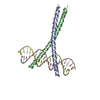
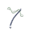
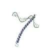
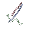
| Title | CRYSTAL STRUCTURE OF C/EBPALPHA-DNA COMPLEX | ||||||
 Components Components |
| ||||||
 Keywords Keywords | TRANSCRIPTION/DNA / BASIC LEUCINE ZIPPER / PROTEIN-DNA COMPLEX / TRANSCRIPTION-DNA COMPLEX | ||||||
| Function / homology |  Function and homology information Function and homology informationnegative regulation of hematopoietic stem cell proliferation / Transcriptional regulation of granulopoiesis / C/EBP complex / CHOP-C/EBP complex / HMG box domain binding / RNA polymerase I transcription regulatory region sequence-specific DNA binding / granulocyte differentiation / urea cycle / myeloid cell differentiation / positive regulation of macrophage activation ...negative regulation of hematopoietic stem cell proliferation / Transcriptional regulation of granulopoiesis / C/EBP complex / CHOP-C/EBP complex / HMG box domain binding / RNA polymerase I transcription regulatory region sequence-specific DNA binding / granulocyte differentiation / urea cycle / myeloid cell differentiation / positive regulation of macrophage activation / epithelial cell maturation / cellular response to lithium ion / interleukin-6-mediated signaling pathway / STAT family protein binding / hematopoietic stem cell proliferation / lipid homeostasis / inner ear development / negative regulation of cell cycle / white fat cell differentiation / macrophage differentiation / positive regulation of osteoblast differentiation / embryonic placenta development / positive regulation of fat cell differentiation / fat cell differentiation / cholesterol metabolic process / transcription by RNA polymerase I / Notch signaling pathway / energy homeostasis / lung development / brown fat cell differentiation / mitochondrion organization / liver development / chromatin DNA binding / cellular response to tumor necrosis factor / kinase binding / positive regulation of inflammatory response / glucose homeostasis / DNA-binding transcription activator activity, RNA polymerase II-specific / sequence-specific DNA binding / DNA-binding transcription factor binding / DNA-binding transcription factor activity, RNA polymerase II-specific / RNA polymerase II cis-regulatory region sequence-specific DNA binding / protein heterodimerization activity / negative regulation of cell population proliferation / negative regulation of DNA-templated transcription / positive regulation of gene expression / regulation of transcription by RNA polymerase II / regulation of DNA-templated transcription / nucleolus / DNA-templated transcription / negative regulation of transcription by RNA polymerase II / protein homodimerization activity / positive regulation of transcription by RNA polymerase II / DNA binding / nucleoplasm / nucleus Similarity search - Function | ||||||
| Biological species |  | ||||||
| Method |  X-RAY DIFFRACTION / SYNCHROTRON / MOLECULAR REPLACEMENT / Resolution: 2.8 Å X-RAY DIFFRACTION / SYNCHROTRON / MOLECULAR REPLACEMENT / Resolution: 2.8 Å | ||||||
 Authors Authors | Miller, M. / Shuman, J.D. / Sebastian, T. / Dauter, Z. / Johnson, P.F. | ||||||
 Citation Citation | Journal: J.Biol.Chem. / Year: 2003 Title: Structural Basis for DNA Recognition by the Basic Region Leucine Zipper Transcription Factor CCAAT/enhancer Binding Protein Alpha Authors: Miller, M. / Shuman, J.D. / Sebastian, T. / Dauter, Z. / Johnson, P.F. | ||||||
| History |
|
- Structure visualization
| Structure viewer | Molecule: MolmilJmol/JSmol |
|---|
- Downloads & links
-Download
| PDBx/mmCIF format | 1nwq.cif.gz | 61.8 KB | Display | PDBx/mmCIF format |
|---|---|---|---|---|
| PDB format | pdb1nwq.ent.gz | 43.4 KB | Display | PDB format |
| PDBx/mmJSON format | 1nwq.json.gz | Tree view | PDBx/mmJSON format | |
| Others |  Other downloads Other downloads |
-Validation report
| Arichive directory | https://data.pdbj.org/pub/pdb/validation_reports/nw/1nwqftp://data.pdbj.org/pub/pdb/validation_reports/nw/1nwq | HTTPS FTP |
|---|
-Related structure data
| Related structure data | 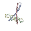 1dh3S S: Starting model for refinement |
|---|---|
| Similar structure data |


-Links
 PDBj
PDBj


- Assembly
| Deposited unit | 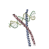
| ||||||||
|---|---|---|---|---|---|---|---|---|---|
| 1 |
| ||||||||
| Unit cell |
|
-Components
| #1: DNA chain | Mass: 6364.120 Da / Num. of mol.: 1 / Source method: obtained synthetically | ||
|---|---|---|---|
| #2: DNA chain | Mass: 6520.249 Da / Num. of mol.: 1 / Source method: obtained synthetically | ||
| #3: Protein | Mass: 7428.297 Da / Num. of mol.: 2 / Fragment: BASIC REGION, LEUCINE ZIPPER DOMAIN Source method: isolated from a genetically manipulated source Source: (gene. exp.)  #4: Water | ChemComp-HOH / |  Mass: 18.015 Da / Num. of mol.: 34 / Source method: isolated from a natural source / Formula: H2O Mass: 18.015 Da / Num. of mol.: 34 / Source method: isolated from a natural source / Formula: H2O |
-Experimental details
-Experiment
| Experiment | Method: X-RAY DIFFRACTION / Number of used crystals: 1 |
|---|
- Sample preparation
| Crystal | Density Matthews: 4.72 Å3/Da / Density % sol: 73.72 % | ||||||||||||||||||||||||||||||||||||||||||||||||||||||||||||||||||||||
|---|---|---|---|---|---|---|---|---|---|---|---|---|---|---|---|---|---|---|---|---|---|---|---|---|---|---|---|---|---|---|---|---|---|---|---|---|---|---|---|---|---|---|---|---|---|---|---|---|---|---|---|---|---|---|---|---|---|---|---|---|---|---|---|---|---|---|---|---|---|---|---|
| Crystal grow | Temperature: 277 K / Method: vapor diffusion, hanging drop / pH: 5.7 Details: NACL, MGCL2, PEG 400, MES, GLYCEROL, pH 5.7, VAPOR DIFFUSION, HANGING DROP, temperature 277K | ||||||||||||||||||||||||||||||||||||||||||||||||||||||||||||||||||||||
| Crystal grow | *PLUS Temperature: 4 ℃ | ||||||||||||||||||||||||||||||||||||||||||||||||||||||||||||||||||||||
| Components of the solutions | *PLUS
|
-Data collection
| Diffraction | Mean temperature: 100 K |
|---|---|
| Diffraction source | Source: SYNCHROTRON / Site: NSLS  / Beamline: X9B / Wavelength: 1.07 Å / Beamline: X9B / Wavelength: 1.07 Å |
| Detector | Type: ADSC QUANTUM 4 / Detector: CCD |
| Radiation | Protocol: SINGLE WAVELENGTH / Monochromatic (M) / Laue (L): M / Scattering type: x-ray |
| Radiation wavelength | Wavelength: 1.07 Å / Relative weight: 1 |
| Reflection | Resolution: 2.8→40 Å / Num. obs: 12416 / % possible obs: 95.6 % / Observed criterion σ(I): -3 / Redundancy: 17.8 % / Biso Wilson estimate: 78.5 Å2 / Rmerge(I) obs: 0.076 / Net I/σ(I): 19.2 |
| Reflection shell | Resolution: 2.8→2.9 Å / Rmerge(I) obs: 0.415 / Mean I/σ(I) obs: 2.7 / % possible all: 91.6 |
| Reflection | *PLUS Highest resolution: 2.8 Å / Lowest resolution: 40 Å / Num. measured all: 222356 |
| Reflection shell | *PLUS Highest resolution: 2.8 Å / Lowest resolution: 2.9 Å / % possible obs: 91.6 % |
- Processing
| Software |
| ||||||||||||||||||||||||||||||||||||||||||||||||||||||||||||||||||||||||||||||||
|---|---|---|---|---|---|---|---|---|---|---|---|---|---|---|---|---|---|---|---|---|---|---|---|---|---|---|---|---|---|---|---|---|---|---|---|---|---|---|---|---|---|---|---|---|---|---|---|---|---|---|---|---|---|---|---|---|---|---|---|---|---|---|---|---|---|---|---|---|---|---|---|---|---|---|---|---|---|---|---|---|---|
| Refinement | Method to determine structure: MOLECULAR REPLACEMENT Starting model: PDB ENTRY 1DH3 Resolution: 2.8→38.53 Å / Rfactor Rfree error: 0.011 / Data cutoff high absF: 1483361.25 / Data cutoff high rms absF: 1483361.25 / Data cutoff low absF: 0 / Isotropic thermal model: RESTRAINED / Cross valid method: THROUGHOUT / σ(F): 0 / Stereochemistry target values: Engh & Huber
| ||||||||||||||||||||||||||||||||||||||||||||||||||||||||||||||||||||||||||||||||
| Solvent computation | Solvent model: FLAT MODEL / Bsol: 37.0305 Å2 / ksol: 0.25376 e/Å3 | ||||||||||||||||||||||||||||||||||||||||||||||||||||||||||||||||||||||||||||||||
| Displacement parameters | Biso mean: 92 Å2
| ||||||||||||||||||||||||||||||||||||||||||||||||||||||||||||||||||||||||||||||||
| Refine analyze |
| ||||||||||||||||||||||||||||||||||||||||||||||||||||||||||||||||||||||||||||||||
| Refinement step | Cycle: LAST / Resolution: 2.8→38.53 Å
| ||||||||||||||||||||||||||||||||||||||||||||||||||||||||||||||||||||||||||||||||
| Refine LS restraints |
| ||||||||||||||||||||||||||||||||||||||||||||||||||||||||||||||||||||||||||||||||
| LS refinement shell | Resolution: 2.8→2.93 Å / Rfactor Rfree error: 0.05 / Total num. of bins used: 8
| ||||||||||||||||||||||||||||||||||||||||||||||||||||||||||||||||||||||||||||||||
| Xplor file |
| ||||||||||||||||||||||||||||||||||||||||||||||||||||||||||||||||||||||||||||||||
| Refinement | *PLUS Highest resolution: 2.8 Å / Lowest resolution: 40 Å | ||||||||||||||||||||||||||||||||||||||||||||||||||||||||||||||||||||||||||||||||
| Solvent computation | *PLUS | ||||||||||||||||||||||||||||||||||||||||||||||||||||||||||||||||||||||||||||||||
| Displacement parameters | *PLUS | ||||||||||||||||||||||||||||||||||||||||||||||||||||||||||||||||||||||||||||||||
| Refine LS restraints | *PLUS
|
