 Movie
Movie Controller

[English] 日本語
 Yorodumi
- PDB-1wz1: Crystal structure of the Fv fragment complexed with dansyl-lysine -
+ Open data

- Basic information
| Entry | Database: PDB / ID: 1wz1 | ||||||
|---|---|---|---|---|---|---|---|
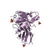
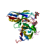
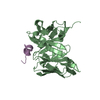
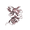
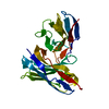
| Title | Crystal structure of the Fv fragment complexed with dansyl-lysine | ||||||
 Components Components |
| ||||||
 Keywords Keywords | IMMUNE SYSTEM / Antigen-antibody fragent complex | ||||||
| Function / homology |  Function and homology information Function and homology informationimmunoglobulin complex / antigen binding / adaptive immune response / immune response / extracellular space Similarity search - Function | ||||||
| Biological species |  | ||||||
| Method |  X-RAY DIFFRACTION / SYNCHROTRON / MOLECULAR REPLACEMENT / Resolution: 1.85 Å X-RAY DIFFRACTION / SYNCHROTRON / MOLECULAR REPLACEMENT / Resolution: 1.85 Å | ||||||
 Authors Authors | Nakasako, M. / Oka, T. / Mashumo, M. / Takahashi, H. / Shimada, I. / Yamaguchi, Y. / Kato, K. / Arata, Y. | ||||||
 Citation Citation | Journal: J.Mol.Biol. / Year: 2005 Title: Conformational dynamics of complementarity-determining region H3 of an anti-dansyl Fv fragment in the presence of its hapten Authors: Nakasako, M. / Oka, T. / Mashumo, M. / Takahashi, H. / Shimada, I. / Yamaguchi, Y. / Kato, K. / Arata, Y. | ||||||
| History |
|
- Structure visualization
| Structure viewer | Molecule: MolmilJmol/JSmol |
|---|
- Downloads & links
-Download
| PDBx/mmCIF format | 1wz1.cif.gz | 59.3 KB | Display | PDBx/mmCIF format |
|---|---|---|---|---|
| PDB format | pdb1wz1.ent.gz | 43.3 KB | Display | PDB format |
| PDBx/mmJSON format | 1wz1.json.gz | Tree view | PDBx/mmJSON format | |
| Others |  Other downloads Other downloads |
-Validation report
| Summary document | 1wz1_validation.pdf.gz | 453.2 KB | Display | wwPDB validaton report |
|---|---|---|---|---|
| Full document | 1wz1_full_validation.pdf.gz | 457.6 KB | Display | |
| Data in XML | 1wz1_validation.xml.gz | 7.1 KB | Display | |
| Data in CIF | 1wz1_validation.cif.gz | 10 KB | Display | |
| Arichive directory | https://data.pdbj.org/pub/pdb/validation_reports/wz/1wz1ftp://data.pdbj.org/pub/pdb/validation_reports/wz/1wz1 | HTTPS FTP |
-Related structure data
| Related structure data |  2dlfS S: Starting model for refinement |
|---|---|
| Similar structure data |


-Links
 PDBj
PDBj

- Assembly
| Deposited unit | 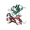
| ||||||||
|---|---|---|---|---|---|---|---|---|---|
| 1 |
| ||||||||
| Unit cell |
|
-Components
| #1: Antibody | Mass: 12386.949 Da / Num. of mol.: 1 Source method: isolated from a genetically manipulated source Source: (gene. exp.)  |
|---|---|
| #2: Antibody | Mass: 13995.593 Da / Num. of mol.: 1 / Fragment: Fv fragment(residues 1-123) Source method: isolated from a genetically manipulated source Source: (gene. exp.) |
| #3: Chemical | ChemComp-DNS /   Type: L-peptide linking / Mass: 379.474 Da / Num. of mol.: 1 / Source method: obtained synthetically / Formula: C18H25N3O4S Type: L-peptide linking / Mass: 379.474 Da / Num. of mol.: 1 / Source method: obtained synthetically / Formula: C18H25N3O4S |
-Experimental details
-Experiment
| Experiment | Method: X-RAY DIFFRACTION / Number of used crystals: 1 |
|---|
- Sample preparation
| Crystal | Density Matthews: 2.35 Å3/Da / Density % sol: 47.66 % |
|---|---|
| Crystal grow | Temperature: 277 K / Method: vapor diffusion, hanging drop / pH: 6.5 Details: PEG 8000, magnecium acetate, sodium cacodylate, pH 6.5, VAPOR DIFFUSION, HANGING DROP, temperature 277K |
-Data collection
| Diffraction | Mean temperature: 110 K |
|---|---|
| Diffraction source | Source: SYNCHROTRON / Site: SPring-8  / Beamline: BL38B1 / Wavelength: 1 Å / Beamline: BL38B1 / Wavelength: 1 Å |
| Detector | Type: RIGAKU RAXIS V / Detector: IMAGE PLATE / Date: Jan 1, 2004 |
| Radiation | Monochromator: Si 111 / Protocol: SINGLE WAVELENGTH / Monochromatic (M) / Laue (L): M / Scattering type: x-ray |
| Radiation wavelength | Wavelength: 1 Å / Relative weight: 1 |
| Reflection | Resolution: 1.8→81.6 Å / Num. obs: 23960 / % possible obs: 100 % / Observed criterion σ(I): 1 / Rmerge(I) obs: 0.078 / Net I/σ(I): 28.4 |
| Reflection shell | Resolution: 1.8→1.82 Å / Rmerge(I) obs: 0.421 / % possible all: 100 |
- Processing
| Software |
| ||||||||||||||||||||
|---|---|---|---|---|---|---|---|---|---|---|---|---|---|---|---|---|---|---|---|---|---|
| Refinement | Method to determine structure: MOLECULAR REPLACEMENT Starting model: PDB ENTRY 2DLF Resolution: 1.85→81.6 Å / Isotropic thermal model: Isotropic / Cross valid method: THROUGHOUT / σ(F): 0 / Stereochemistry target values: Engh & Huber
| ||||||||||||||||||||
| Refinement step | Cycle: LAST / Resolution: 1.85→81.6 Å
| ||||||||||||||||||||
| Refine LS restraints |
|
