 Movie
Movie Controller


+ Open data

- Basic information
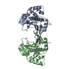
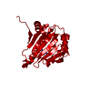
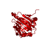
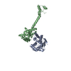
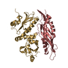
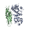
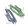
| Entry | Database: PDB / ID: 1usu | ||||||
|---|---|---|---|---|---|---|---|
| Title | The Structure of the complex between Aha1 and HSP90 | ||||||
 Components Components |
| ||||||
 Keywords Keywords | CHAPERONE / CHAPERONE-COMPLEX / ACTIVATOR / HSP90 | ||||||
| Function / homology |  Function and homology information Function and homology informationThe NLRP3 inflammasome / Tetrahydrobiopterin (BH4) synthesis, recycling, salvage and regulation / eNOS activation / Extra-nuclear estrogen signaling / VEGFR2 mediated vascular permeability / HSF1-dependent transactivation / HSP90 chaperone cycle for steroid hormone receptors (SHR) in the presence of ligand / HSF1 activation / response to oxygen levels / box C/D snoRNP assembly ...The NLRP3 inflammasome / Tetrahydrobiopterin (BH4) synthesis, recycling, salvage and regulation / eNOS activation / Extra-nuclear estrogen signaling / VEGFR2 mediated vascular permeability / HSF1-dependent transactivation / HSP90 chaperone cycle for steroid hormone receptors (SHR) in the presence of ligand / HSF1 activation / response to oxygen levels / box C/D snoRNP assembly / response to osmotic stress / regulation of telomere maintenance / 'de novo' protein folding / : / ATPase activator activity / proteasome assembly / positive regulation of telomere maintenance via telomerase / Neutrophil degranulation / protein maturation / ATP-dependent protein folding chaperone / protein import into nucleus / unfolded protein binding / protein-folding chaperone binding / cellular response to heat / protein folding / protein refolding / protein stabilization / perinuclear region of cytoplasm / ATP hydrolysis activity / protein-containing complex / ATP binding / identical protein binding / nucleus / plasma membrane / cytoplasm / cytosol Similarity search - Function | ||||||
| Biological species |  | ||||||
| Method |  X-RAY DIFFRACTION / SYNCHROTRON / MOLECULAR REPLACEMENT / Resolution: 2.15 Å X-RAY DIFFRACTION / SYNCHROTRON / MOLECULAR REPLACEMENT / Resolution: 2.15 Å | ||||||
 Authors Authors | Meyer, P. / Roe, S.M. / Pearl, L.H. | ||||||
 Citation Citation | Journal: Embo J. / Year: 2004 Title: Structural Basis for Recruitment of the ATPase Activator Aha1 to the Hsp90 Chaperone Machinery. Authors: Meyer, P. / Prodromou, C. / Liao, C. / Hu, B. / Roe, S.M. / Vaughan, C.K. / Vlasic, I. / Panaretou, B. / Piper, P.W. / Pearl, L.H. | ||||||
| History |
| ||||||
| Remark 700 | SHEET THE SHEET STRUCTURE OF THIS MOLECULE IS BIFURCATED. IN ORDER TO REPRESENT THIS FEATURE IN ... SHEET THE SHEET STRUCTURE OF THIS MOLECULE IS BIFURCATED. IN ORDER TO REPRESENT THIS FEATURE IN THE SHEET RECORDS BELOW, TWO SHEETS ARE DEFINED. |
- Structure visualization
| Structure viewer | Molecule: MolmilJmol/JSmol |
|---|
- Downloads & links
-Download
| PDBx/mmCIF format | 1usu.cif.gz | 97.4 KB | Display | PDBx/mmCIF format |
|---|---|---|---|---|
| PDB format | pdb1usu.ent.gz | 72.5 KB | Display | PDB format |
| PDBx/mmJSON format | 1usu.json.gz | Tree view | PDBx/mmJSON format | |
| Others |  Other downloads Other downloads |
-Validation report
| Arichive directory | https://data.pdbj.org/pub/pdb/validation_reports/us/1usuftp://data.pdbj.org/pub/pdb/validation_reports/us/1usu | HTTPS FTP |
|---|
-Related structure data
| Related structure data |  1usvC 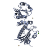 1hk7S S: Starting model for refinement C: citing same article ( |
|---|---|
| Similar structure data |


-Links
 PDBj
PDBj


- Assembly
| Deposited unit | 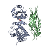
| ||||||||
|---|---|---|---|---|---|---|---|---|---|
| 1 |
| ||||||||
| Unit cell |
|
-Components
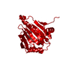
| #1: Protein | Mass: 30486.727 Da / Num. of mol.: 1 / Fragment: MIDDLE DOMAIN, RESIDUES 273-530 Source method: isolated from a genetically manipulated source Source: (gene. exp.) Production host:  |
|---|---|
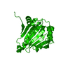
| #2: Protein | Mass: 19118.715 Da / Num. of mol.: 1 / Fragment: N-TERMINAL DOMAIN, RESIDUES 1-156 Source method: isolated from a genetically manipulated source Source: (gene. exp.) Production host: |
| #3: Chemical | ChemComp-GOL /   Mass: 92.094 Da / Num. of mol.: 1 / Source method: obtained synthetically / Formula: C3H8O3 Mass: 92.094 Da / Num. of mol.: 1 / Source method: obtained synthetically / Formula: C3H8O3 |
| #4: Water | ChemComp-HOH /  Mass: 18.015 Da / Num. of mol.: 227 / Source method: isolated from a natural source / Formula: H2O Mass: 18.015 Da / Num. of mol.: 227 / Source method: isolated from a natural source / Formula: H2O |
-Experimental details
-Experiment
| Experiment | Method: X-RAY DIFFRACTION / Number of used crystals: 1 |
|---|
- Sample preparation
| Crystal | Density Matthews: 2.6 Å3/Da / Density % sol: 51.6 % | ||||||||||||||||||||||||||||||||||||||||||||||||||||||||
|---|---|---|---|---|---|---|---|---|---|---|---|---|---|---|---|---|---|---|---|---|---|---|---|---|---|---|---|---|---|---|---|---|---|---|---|---|---|---|---|---|---|---|---|---|---|---|---|---|---|---|---|---|---|---|---|---|---|
| Crystal grow | Temperature: 292 K / Method: microbatch / pH: 6.5 Details: CRYSTALS GREW FROM A MIXTURE OF MIDDLE DOMAIN HSP90 AND N- TERMINAL AHA1 AT A FINAL CONCENTRATION OF 110 UM AND 165 UM, RESPECTIVELY, IN A SOLUTION CONTAINING 90 MM AMMONIUM SULPHATE, 13.5% ...Details: CRYSTALS GREW FROM A MIXTURE OF MIDDLE DOMAIN HSP90 AND N- TERMINAL AHA1 AT A FINAL CONCENTRATION OF 110 UM AND 165 UM, RESPECTIVELY, IN A SOLUTION CONTAINING 90 MM AMMONIUM SULPHATE, 13.5% (W/V) PEG8K AND 45 MM SODIUM CACODYLATE PH 6.5 IN UNDER-OIL MICROBATCH EXPERIMENTS AT 19C. | ||||||||||||||||||||||||||||||||||||||||||||||||||||||||
| Crystal grow | *PLUS Temperature: 19 ℃ / pH: 6.5 / Method: batch method | ||||||||||||||||||||||||||||||||||||||||||||||||||||||||
| Components of the solutions | *PLUS
|
-Data collection
| Diffraction | Mean temperature: 100 K |
|---|---|
| Diffraction source | Source: SYNCHROTRON / Site: ESRF  / Beamline: ID14-1 / Wavelength: 0.934 / Beamline: ID14-1 / Wavelength: 0.934 |
| Detector | Type: ADSC CCD / Detector: CCD / Date: Nov 15, 2002 |
| Radiation | Protocol: SINGLE WAVELENGTH / Monochromatic (M) / Laue (L): M / Scattering type: x-ray |
| Radiation wavelength | Wavelength: 0.934 Å / Relative weight: 1 |
| Reflection | Resolution: 2.15→50 Å / Num. obs: 26832 / % possible obs: 99.3 % / Redundancy: 3.9 % / Biso Wilson estimate: 31.1 Å2 / Rmerge(I) obs: 0.062 / Net I/σ(I): 6.6 |
| Reflection shell | Resolution: 2.15→2.27 Å / Redundancy: 3.7 % / Rmerge(I) obs: 0.218 / Mean I/σ(I) obs: 3.2 / % possible all: 99.4 |
| Reflection | *PLUS Highest resolution: 2.15 Å / % possible obs: 99.1 % / Redundancy: 3.6 % / Rmerge F obs: 0.089 |
| Reflection shell | *PLUS % possible obs: 99 % / Rmerge(I) obs: 0.302 / Mean I/σ(I) obs: 2.3 |
- Processing
| Software |
| ||||||||||||||||||||||||||||||||||||||||||||||||||||||||||||||||||||||||||||||||
|---|---|---|---|---|---|---|---|---|---|---|---|---|---|---|---|---|---|---|---|---|---|---|---|---|---|---|---|---|---|---|---|---|---|---|---|---|---|---|---|---|---|---|---|---|---|---|---|---|---|---|---|---|---|---|---|---|---|---|---|---|---|---|---|---|---|---|---|---|---|---|---|---|---|---|---|---|---|---|---|---|---|
| Refinement | Method to determine structure: MOLECULAR REPLACEMENT Starting model: PDB ENTRY 1HK7 Resolution: 2.15→31.79 Å / Rfactor Rfree error: 0.007 / Data cutoff high absF: 1001963.23 / Isotropic thermal model: RESTRAINED / Cross valid method: THROUGHOUT / σ(F): 0
| ||||||||||||||||||||||||||||||||||||||||||||||||||||||||||||||||||||||||||||||||
| Solvent computation | Solvent model: FLAT MODEL / Bsol: 50.5145 Å2 / ksol: 0.340679 e/Å3 | ||||||||||||||||||||||||||||||||||||||||||||||||||||||||||||||||||||||||||||||||
| Displacement parameters | Biso mean: 48.8 Å2
| ||||||||||||||||||||||||||||||||||||||||||||||||||||||||||||||||||||||||||||||||
| Refine analyze |
| ||||||||||||||||||||||||||||||||||||||||||||||||||||||||||||||||||||||||||||||||
| Refinement step | Cycle: LAST / Resolution: 2.15→31.79 Å
| ||||||||||||||||||||||||||||||||||||||||||||||||||||||||||||||||||||||||||||||||
| Refine LS restraints |
| ||||||||||||||||||||||||||||||||||||||||||||||||||||||||||||||||||||||||||||||||
| LS refinement shell | Resolution: 2.15→2.28 Å / Rfactor Rfree error: 0.02 / Total num. of bins used: 6
| ||||||||||||||||||||||||||||||||||||||||||||||||||||||||||||||||||||||||||||||||
| Xplor file |
| ||||||||||||||||||||||||||||||||||||||||||||||||||||||||||||||||||||||||||||||||
| Refinement | *PLUS Lowest resolution: 100 Å / Rfactor Rwork: 0.214 | ||||||||||||||||||||||||||||||||||||||||||||||||||||||||||||||||||||||||||||||||
| Solvent computation | *PLUS | ||||||||||||||||||||||||||||||||||||||||||||||||||||||||||||||||||||||||||||||||
| Displacement parameters | *PLUS | ||||||||||||||||||||||||||||||||||||||||||||||||||||||||||||||||||||||||||||||||
| Refine LS restraints | *PLUS
|
