 Movie
Movie Controller


+ Open data

- Basic information
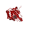
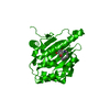
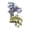
| Entry | Database: PDB / ID: 1amw | ||||||
|---|---|---|---|---|---|---|---|
| Title | ADP BINDING SITE IN THE HSP90 MOLECULAR CHAPERONE | ||||||
 Components Components | HEAT SHOCK PROTEIN 90 | ||||||
 Keywords Keywords | CHAPERONE / NUCLEOTIDE BINDING SITE | ||||||
| Function / homology |  Function and homology information Function and homology informationThe NLRP3 inflammasome / Tetrahydrobiopterin (BH4) synthesis, recycling, salvage and regulation / eNOS activation / Extra-nuclear estrogen signaling / VEGFR2 mediated vascular permeability / HSP90 chaperone cycle for steroid hormone receptors (SHR) in the presence of ligand / HSF1-dependent transactivation / HSF1 activation / response to oxygen levels / box C/D snoRNP assembly ...The NLRP3 inflammasome / Tetrahydrobiopterin (BH4) synthesis, recycling, salvage and regulation / eNOS activation / Extra-nuclear estrogen signaling / VEGFR2 mediated vascular permeability / HSP90 chaperone cycle for steroid hormone receptors (SHR) in the presence of ligand / HSF1-dependent transactivation / HSF1 activation / response to oxygen levels / box C/D snoRNP assembly / : / response to osmotic stress / regulation of telomere maintenance / 'de novo' protein folding / proteasome assembly / positive regulation of telomere maintenance via telomerase / Neutrophil degranulation / ATP-dependent protein folding chaperone / protein maturation / : / cellular response to heat / protein refolding / protein folding / protein stabilization / perinuclear region of cytoplasm / ATP hydrolysis activity / protein-containing complex / ATP binding / identical protein binding / nucleus / plasma membrane / cytosol / cytoplasm Similarity search - Function | ||||||
| Biological species |  | ||||||
| Method |  X-RAY DIFFRACTION / SYNCHROTRON / ISOMORPHOUS REPLACEMENT / Resolution: 1.85 Å X-RAY DIFFRACTION / SYNCHROTRON / ISOMORPHOUS REPLACEMENT / Resolution: 1.85 Å | ||||||
 Authors Authors | Pearl, L.H. / Roe, S.M. / Prodromou, C. | ||||||
 Citation Citation | Journal: Cell(Cambridge,Mass.) / Year: 1997 Title: Identification and structural characterization of the ATP/ADP-binding site in the Hsp90 molecular chaperone Authors: Prodromou, C. / Roe, S.M. / O'Brien, R. / Ladbury, J.E. / Piper, P.W. / Pearl, L.H. #1: Journal: Nat.Struct.Biol. / Year: 1997Title: A Molecular Clamp in the Crystal Structure of the N-Terminal Domain of the Yeast Hsp90 Chaperone Authors: Prodromou, C. / Roe, S.M. / Piper, P.W. / Pearl, L.H. | ||||||
| History |
|
- Structure visualization
| Structure viewer | Molecule: MolmilJmol/JSmol |
|---|
- Downloads & links
-Download
| PDBx/mmCIF format | 1amw.cif.gz | 64.3 KB | Display | PDBx/mmCIF format |
|---|---|---|---|---|
| PDB format | pdb1amw.ent.gz | 46.4 KB | Display | PDB format |
| PDBx/mmJSON format | 1amw.json.gz | Tree view | PDBx/mmJSON format | |
| Others |  Other downloads Other downloads |
-Validation report
| Arichive directory | https://data.pdbj.org/pub/pdb/validation_reports/am/1amwftp://data.pdbj.org/pub/pdb/validation_reports/am/1amw | HTTPS FTP |
|---|
-Related structure data
| Related structure data | 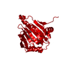 1a4hC 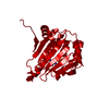 1am1C 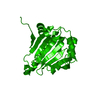 1ah6S S: Starting model for refinement C: citing same article ( |
|---|---|
| Similar structure data |


-Links
 PDBj
PDBj


- Assembly
| Deposited unit | 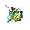
| ||||||||
|---|---|---|---|---|---|---|---|---|---|
| 1 | 
| ||||||||
| Unit cell |
| ||||||||
| Components on special symmetry positions |
|
-Components
| #1: Protein | Mass: 24208.582 Da / Num. of mol.: 1 / Fragment: N-TERMINAL RESIDUES Source method: isolated from a genetically manipulated source Details: ADP COMPLEX Source: (gene. exp.) Plasmid: PRSETA / Production host:  |
|---|---|
| #2: Chemical | ChemComp-ADP /   Mass: 427.201 Da / Num. of mol.: 1 / Source method: obtained synthetically / Formula: C10H15N5O10P2 / Comment: ADP, energy-carrying molecule*YM Mass: 427.201 Da / Num. of mol.: 1 / Source method: obtained synthetically / Formula: C10H15N5O10P2 / Comment: ADP, energy-carrying molecule*YM |
| #3: Water | ChemComp-HOH /  Mass: 18.015 Da / Num. of mol.: 344 / Source method: isolated from a natural source / Formula: H2O Mass: 18.015 Da / Num. of mol.: 344 / Source method: isolated from a natural source / Formula: H2O |
-Experimental details
-Experiment
| Experiment | Method: X-RAY DIFFRACTION / Number of used crystals: 1 |
|---|
- Sample preparation
| Crystal | Density Matthews: 3.2 Å3/Da / Density % sol: 61 % | ||||||||||||||||||||||||||||||||||||||||||
|---|---|---|---|---|---|---|---|---|---|---|---|---|---|---|---|---|---|---|---|---|---|---|---|---|---|---|---|---|---|---|---|---|---|---|---|---|---|---|---|---|---|---|---|
| Crystal grow | Method: under oil / pH: 5 Details: PROTEIN WAS CRYSTALLIZED UNDER OIL IN TERASAKI PLATES. THE DROPS CONTAINED 27MG/ML PROTEIN, 9.75%(W/V) PEGME 550, 65MM AMMONIUM SULFATE, 32.5MM SODIUM SUCCINATE PH5.0, 5MM ADP AND 5MM ...Details: PROTEIN WAS CRYSTALLIZED UNDER OIL IN TERASAKI PLATES. THE DROPS CONTAINED 27MG/ML PROTEIN, 9.75%(W/V) PEGME 550, 65MM AMMONIUM SULFATE, 32.5MM SODIUM SUCCINATE PH5.0, 5MM ADP AND 5MM MAGNESIUM CHLORIDE., under oil | ||||||||||||||||||||||||||||||||||||||||||
| Crystal grow | *PLUS Method: unknown | ||||||||||||||||||||||||||||||||||||||||||
| Components of the solutions | *PLUS
|
-Data collection
| Diffraction | Mean temperature: 110 K |
|---|---|
| Diffraction source | Source: SYNCHROTRON / Site: SRS  / Beamline: PX9.5 / Wavelength: 0.92 / Beamline: PX9.5 / Wavelength: 0.92 |
| Detector | Type: MARRESEARCH / Detector: IMAGE PLATE / Date: Jun 1, 1997 / Details: TORROIDAL PT-COATED SI MIRROR |
| Radiation | Monochromator: SI(111) / Monochromatic (M) / Laue (L): M / Scattering type: x-ray |
| Radiation wavelength | Wavelength: 0.92 Å / Relative weight: 1 |
| Reflection | Resolution: 1.84→24 Å / Num. obs: 27146 / % possible obs: 99 % / Observed criterion σ(I): 2 / Redundancy: 3.5 % / Biso Wilson estimate: 11.6 Å2 / Rsym value: 0.093 / Net I/σ(I): 4.6 |
| Reflection shell | Resolution: 1.84→1.89 Å / Redundancy: 3.4 % / Mean I/σ(I) obs: 3.2 / Rsym value: 0.228 / % possible all: 93.7 |
| Reflection | *PLUS Rmerge(I) obs: 0.093 |
| Reflection shell | *PLUS % possible obs: 93.7 % / Rmerge(I) obs: 0.228 |
- Processing
| Software |
| ||||||||||||||||||||||||||||||||||||||||||||||||||||||||||||
|---|---|---|---|---|---|---|---|---|---|---|---|---|---|---|---|---|---|---|---|---|---|---|---|---|---|---|---|---|---|---|---|---|---|---|---|---|---|---|---|---|---|---|---|---|---|---|---|---|---|---|---|---|---|---|---|---|---|---|---|---|---|
| Refinement | Method to determine structure: ISOMORPHOUS REPLACEMENT Starting model: PDB ENTRY 1AH6 Resolution: 1.85→8 Å / Data cutoff high absF: 1000000 / Data cutoff low absF: 0.001 / Isotropic thermal model: RESTRAINED / σ(F): 2 / Details: BULK SOLVENT MODEL USED
| ||||||||||||||||||||||||||||||||||||||||||||||||||||||||||||
| Displacement parameters | Biso mean: 20.4 Å2 | ||||||||||||||||||||||||||||||||||||||||||||||||||||||||||||
| Refine analyze | Luzzati coordinate error obs: 0.18 Å / Luzzati d res low obs: 5 Å / Luzzati sigma a obs: 0.16 Å | ||||||||||||||||||||||||||||||||||||||||||||||||||||||||||||
| Refinement step | Cycle: LAST / Resolution: 1.85→8 Å
| ||||||||||||||||||||||||||||||||||||||||||||||||||||||||||||
| Refine LS restraints |
| ||||||||||||||||||||||||||||||||||||||||||||||||||||||||||||
| LS refinement shell | Resolution: 1.85→1.93 Å / Total num. of bins used: 8
| ||||||||||||||||||||||||||||||||||||||||||||||||||||||||||||
| Xplor file |
| ||||||||||||||||||||||||||||||||||||||||||||||||||||||||||||
| Software | *PLUS Name: X-PLOR / Version: 3.851 / Classification: refinement | ||||||||||||||||||||||||||||||||||||||||||||||||||||||||||||
| Refinement | *PLUS Rfactor Rfree: 0.243 | ||||||||||||||||||||||||||||||||||||||||||||||||||||||||||||
| Solvent computation | *PLUS | ||||||||||||||||||||||||||||||||||||||||||||||||||||||||||||
| Displacement parameters | *PLUS | ||||||||||||||||||||||||||||||||||||||||||||||||||||||||||||
| Refine LS restraints | *PLUS
| ||||||||||||||||||||||||||||||||||||||||||||||||||||||||||||
| LS refinement shell | *PLUS Rfactor obs: 0.223 |
