 Movie
Movie Controller

[English] 日本語
 Yorodumi
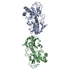
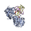
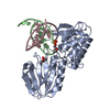
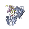
- PDB-1pjj: Complex between the Lactococcus lactis Fpg and an abasic site con... -
+ Open data

- Basic information
| Entry | Database: PDB / ID: 1pjj | ||||||
|---|---|---|---|---|---|---|---|
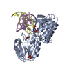
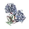
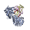
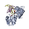
| Title | Complex between the Lactococcus lactis Fpg and an abasic site containing DNA. | ||||||
 Components Components |
| ||||||
 Keywords Keywords | hydrolase/DNA /  DNA repair / Fpg / MutM / abasic site / hydrolase-DNA COMPLEX DNA repair / Fpg / MutM / abasic site / hydrolase-DNA COMPLEX | ||||||
| Function / homology |  Function and homology informationDNA-formamidopyrimidine glycosylase / oxidized purine nucleobase lesion DNA N-glycosylase activity / class I DNA-(apurinic or apyrimidinic site) endonuclease activity / DNA-(apurinic or apyrimidinic site) lyase / base-excision repair / damaged DNA binding / zinc ion binding Function and homology informationDNA-formamidopyrimidine glycosylase / oxidized purine nucleobase lesion DNA N-glycosylase activity / class I DNA-(apurinic or apyrimidinic site) endonuclease activity / DNA-(apurinic or apyrimidinic site) lyase / base-excision repair / damaged DNA binding / zinc ion bindingSimilarity search - Function | ||||||
| Biological species |  Lactococcus lactis (lactic acid bacteria) Lactococcus lactis (lactic acid bacteria) | ||||||
| Method | X-RAY DIFFRACTION / SYNCHROTRON / MOLECULAR REPLACEMENT / Resolution: 1.9 Å | ||||||
 Authors Authors | Serre, L. / Pereira de Jesus, K. / Boiteux, S. / Zelwer, C. / Castaing, B. | ||||||
 Citation Citation | Journal: Nucleic Acids Res. / Year: 2005 Title: Structural insights into abasic site for Fpg specific binding and catalysis: comparative high-resolution crystallographic studies of Fpg bound to various models of abasic site analogues-containing DNA. Authors: Pereira de Jesus, K. / Serre, L. / Zelwer, C. / Castaing, B. #1: Journal: Acta Crystallogr.,Sect.D / Year: 2002Title: Crystallization and primary X-ray crystallographic studies of a complex between th Lactococcus lactis Fpg DNA- repair enzyme and an abasic site containing DNA Authors: Pereira de Jesus, K. / Serre, L. / Hervouet, N. / Bouckson-Castaing, V. / Zelwer, C. / Castaing, B. | ||||||
| History |
| ||||||
| Remark 999 | sequence The author maintains that the Asp137 is an error in the sequence database. This residue ...sequence The author maintains that the Asp137 is an error in the sequence database. This residue does not exist. |
- Structure visualization
| Structure viewer | Molecule: MolmilJmol/JSmol |
|---|
- Downloads & links
-Download
| PDBx/mmCIF format | 1pjj.cif.gz | 92.8 KB | Display | PDBx/mmCIF format |
|---|---|---|---|---|
| PDB format | pdb1pjj.ent.gz | 66.1 KB | Display | PDB format |
| PDBx/mmJSON format | 1pjj.json.gz | Tree view | PDBx/mmJSON format | |
| Others |  Other downloads Other downloads |
-Validation report
| Arichive directory | https://data.pdbj.org/pub/pdb/validation_reports/pj/1pjjftp://data.pdbj.org/pub/pdb/validation_reports/pj/1pjj | HTTPS FTP |
|---|
-Related structure data
| Related structure data |  1nnjC  1pjiC  1pm5C  1kfvS S: Starting model for refinement C: citing same article ( |
|---|---|
| Similar structure data |


-Links
 PDBj
PDBj


- Assembly
| Deposited unit | 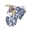
| ||||||||
|---|---|---|---|---|---|---|---|---|---|
| 1 |
| ||||||||
| Unit cell |
|
-Components
-DNA chain , 2 types, 2 molecules DE
| #1: DNA chain | Mass: 4054.614 Da / Num. of mol.: 1 / Source method: obtained synthetically |
|---|---|
| #2: DNA chain | Mass: 4355.884 Da / Num. of mol.: 1 / Source method: obtained synthetically |
-Protein , 1 types, 1 molecules A
| #3: Protein | DNA-formamidopyrimidine glycosylase / 3.2.2.23 / FAPY-DNA glycosylase Mass: 31076.154 Da / Num. of mol.: 1 / Mutation: P1G Source method: isolated from a genetically manipulated source Source: (gene. exp.) Lactococcus lactis (lactic acid bacteria)Gene: MUTM OR FPG / Plasmid: pmal-c / Production host: Escherichia coli (E. coli)References: UniProt: P42371, DNA-formamidopyrimidine glycosylase |
|---|
-Non-polymers , 3 types, 282 molecules 


| #4: Chemical | ChemComp-ZN /  Mass: 65.409 Da / Num. of mol.: 1 / Source method: obtained synthetically / Formula: Zn Mass: 65.409 Da / Num. of mol.: 1 / Source method: obtained synthetically / Formula: Zn | ||
|---|---|---|---|
| #5: Chemical | Glycerol Mass: 92.094 Da / Num. of mol.: 3 / Source method: obtained synthetically / Formula: C3H8O3 Mass: 92.094 Da / Num. of mol.: 3 / Source method: obtained synthetically / Formula: C3H8O3#6: Water | ChemComp-HOH / | WaterMass: 18.015 Da / Num. of mol.: 278 / Source method: isolated from a natural source / Formula: H2O |
-Experimental details
-Experiment
| Experiment | Method: X-RAY DIFFRACTION / Number of used crystals: 1 |
|---|
- Sample preparation
| Crystal | Density Matthews: 3.78 Å3/Da / Density % sol: 67.43 % | ||||||||||||||||||||||||||||
|---|---|---|---|---|---|---|---|---|---|---|---|---|---|---|---|---|---|---|---|---|---|---|---|---|---|---|---|---|---|
| Crystal grow | Temperature: 298 K / Method: vapor diffusion, hanging drop / pH: 8 Details: citrate, Hepes, glycerol, pH 8.0, VAPOR DIFFUSION, HANGING DROP, temperature 298K | ||||||||||||||||||||||||||||
| Components of the solutions |
|
-Data collection
| Diffraction | Mean temperature: 100 K |
|---|---|
| Diffraction source | Source: SYNCHROTRON / Site: ESRF  / Beamline: ID14-2 / Wavelength: 0.93 Å / Beamline: ID14-2 / Wavelength: 0.93 Å |
| Detector | Type: ADSC QUANTUM 4 / Detector: CCD / Date: Jul 4, 2002 |
| Radiation | Monochromator: Mirror / Protocol: SINGLE WAVELENGTH / Monochromatic (M) / Laue (L): M / Scattering type: x-ray |
| Radiation wavelength | Wavelength: 0.93 Å / Relative weight: 1 |
| Reflection | Resolution: 1.9→43.7 Å / Num. all: 48837 / Num. obs: 48787 / % possible obs: 100 % / Redundancy: 9.6 % / Biso Wilson estimate: 27.689 Å2 / Rmerge(I) obs: 0.077 / Rsym value: 0.073 / Net I/σ(I): 5.2 |
| Reflection shell | Resolution: 1.9→2 Å / Redundancy: 9.7 % / Rmerge(I) obs: 0.516 / Mean I/σ(I) obs: 0.8 / Num. unique all: 7019 / Rsym value: 0.488 / % possible all: 100 |
- Processing
| Software |
| |||||||||||||||||||||||||
|---|---|---|---|---|---|---|---|---|---|---|---|---|---|---|---|---|---|---|---|---|---|---|---|---|---|---|
| Refinement | Method to determine structure: MOLECULAR REPLACEMENT Starting model: PDB ENTRY 1KFV Resolution: 1.9→35 Å / Isotropic thermal model: Isotropic / Cross valid method: THROUGHOUT / σ(F): 0 / Stereochemistry target values: Engh & Huber
| |||||||||||||||||||||||||
| Displacement parameters | Biso mean: 31 Å2 | |||||||||||||||||||||||||
| Refinement step | Cycle: LAST / Resolution: 1.9→35 Å
| |||||||||||||||||||||||||
| LS refinement shell | Resolution: 1.9→1.91 Å /
|
