 Movie
Movie Controller

[English] 日本語
 Yorodumi
- PDB-1p6a: STRUCTURAL BASIS FOR VARIATION IN ADENOVIRUS AFFINITY FOR THE CEL... -
+ Open data

- Basic information
| Entry | Database: PDB / ID: 1p6a | ||||||
|---|---|---|---|---|---|---|---|
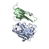
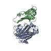
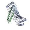
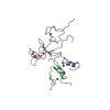
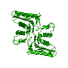
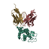
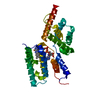
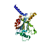
| Title | STRUCTURAL BASIS FOR VARIATION IN ADENOVIRUS AFFINITY FOR THE CELLULAR RECEPTOR CAR (S489Y MUTANT) | ||||||
 Components Components |
| ||||||
 Keywords Keywords | Viral protein/receptor / VIRUS / VIRAL PROTEIN / Viral protein-receptor COMPLEX | ||||||
| Function / homology |  Function and homology information Function and homology informationAV node cell-bundle of His cell adhesion involved in cell communication / cell adhesive protein binding involved in AV node cell-bundle of His cell communication / AV node cell to bundle of His cell communication / homotypic cell-cell adhesion / epithelial structure maintenance / regulation of AV node cell action potential / gamma-delta T cell activation / apicolateral plasma membrane / germ cell migration / connexin binding ...AV node cell-bundle of His cell adhesion involved in cell communication / cell adhesive protein binding involved in AV node cell-bundle of His cell communication / AV node cell to bundle of His cell communication / homotypic cell-cell adhesion / epithelial structure maintenance / regulation of AV node cell action potential / gamma-delta T cell activation / apicolateral plasma membrane / germ cell migration / connexin binding / transepithelial transport / cell-cell junction organization / adhesion receptor-mediated virion attachment to host cell / cardiac muscle cell development / heterophilic cell-cell adhesion / intercalated disc / bicellular tight junction / neutrophil chemotaxis / cell adhesion molecule binding / acrosomal vesicle / Cell surface interactions at the vascular wall / PDZ domain binding / adherens junction / filopodium / mitochondrion organization / neuromuscular junction / beta-catenin binding / integrin binding / Immunoregulatory interactions between a Lymphoid and a non-Lymphoid cell / cell-cell junction / viral capsid / cell junction / heart development / growth cone / cell body / virus receptor activity / actin cytoskeleton organization / basolateral plasma membrane / defense response to virus / cell adhesion / neuron projection / membrane raft / signaling receptor binding / symbiont entry into host cell / host cell nucleus / protein-containing complex / extracellular space / extracellular region / nucleoplasm / identical protein binding / plasma membrane / cytoplasm Similarity search - Function | ||||||
| Biological species |  Human adenovirus A serotype 12 Human adenovirus A serotype 12 Homo sapiens (human) Homo sapiens (human) | ||||||
| Method |  X-RAY DIFFRACTION / SYNCHROTRON / FOURIER SYNTHESIS / Resolution: 2.9 Å X-RAY DIFFRACTION / SYNCHROTRON / FOURIER SYNTHESIS / Resolution: 2.9 Å | ||||||
 Authors Authors | Howitt, J. / Bewley, M.C. / Graziano, V. / Flanagan, J.M. / Freimuth, P. | ||||||
 Citation Citation | Journal: J.Biol.Chem. / Year: 2003 Title: Structural basis for variation in adenovirus affinity for the cellular coxsackievirus and adenovirus receptor. Authors: Howitt, J. / Bewley, M.C. / Graziano, V. / Flanagan, J.M. / Freimuth, P. #1: Journal: Science / Year: 1999Title: Structural analysis of the mechanism of adenovirus binding to its human cellular receptor, CAR Authors: Bewley, M.C. / Springer, K. / Zhang, Y.B. / Freimuth, P. / Flanagan, J.M. #2: Journal: J.Virol. / Year: 1999Title: Coxsackievirus and adenovirus receptor amino-terminal immunoglobulin V-related domain binds adenovirus type 2 and fiber knob from adenovirus type 12 Authors: Freimuth, P. / Springer, K. / Berard, C. / Hainfeld, J. / Bewley, M.C. / Flanagan, J.M. | ||||||
| History |
|
- Structure visualization
| Structure viewer | Molecule: MolmilJmol/JSmol |
|---|
- Downloads & links
-Download
| PDBx/mmCIF format | 1p6a.cif.gz | 70.7 KB | Display | PDBx/mmCIF format |
|---|---|---|---|---|
| PDB format | pdb1p6a.ent.gz | 53.2 KB | Display | PDB format |
| PDBx/mmJSON format | 1p6a.json.gz | Tree view | PDBx/mmJSON format | |
| Others |  Other downloads Other downloads |
-Validation report
| Arichive directory | https://data.pdbj.org/pub/pdb/validation_reports/p6/1p6aftp://data.pdbj.org/pub/pdb/validation_reports/p6/1p6a | HTTPS FTP |
|---|
-Related structure data


-Links
 PDBj
PDBj


- Assembly
| Deposited unit | 
| ||||||||
|---|---|---|---|---|---|---|---|---|---|
| 1 |
| ||||||||
| Unit cell |
|
-Components
| #1: Protein | Mass: 20020.572 Da / Num. of mol.: 1 / Fragment: knob domain (UNP residues 403-587) / Mutation: Y489S Source method: isolated from a genetically manipulated source Source: (gene. exp.) Human adenovirus A serotype 12 / Genus: Mastadenovirus / Species: Human adenovirus A / Gene: L5 / Plasmid: pET15b / Production host:  |
|---|---|
| #2: Protein | Mass: 13640.500 Da / Num. of mol.: 1 Source method: isolated from a genetically manipulated source Source: (gene. exp.) Homo sapiens (human) / Gene: CXADR, CAR / Plasmid: pET15b / Production host: |
| Has protein modification | Y |
-Experimental details
-Experiment
| Experiment | Method: X-RAY DIFFRACTION |
|---|
- Sample preparation
| Crystal | Density Matthews: 5.97 Å3/Da / Density % sol: 79.39 % |
|---|---|
| Crystal grow | Method: vapor diffusion, hanging drop / Details: VAPOR DIFFUSION, HANGING DROP |
-Data collection
| Diffraction | Mean temperature: 99 K |
|---|---|
| Diffraction source | Source: SYNCHROTRON / Site: NSLS  / Beamline: X25 / Wavelength: 1 Å / Beamline: X25 / Wavelength: 1 Å |
| Detector | Type: CUSTOM-MADE / Detector: CCD |
| Radiation | Protocol: SINGLE WAVELENGTH / Monochromatic (M) / Laue (L): M / Scattering type: x-ray |
| Radiation wavelength | Wavelength: 1 Å / Relative weight: 1 |
| Reflection | Resolution: 2.9→20 Å |
- Processing
| Software |
| ||||||||||||
|---|---|---|---|---|---|---|---|---|---|---|---|---|---|
| Refinement | Method to determine structure: FOURIER SYNTHESIS / Resolution: 2.9→20 Å / Cross valid method: THROUGHOUT / Stereochemistry target values: Engh & Huber /
| ||||||||||||
| Refinement step | Cycle: LAST / Resolution: 2.9→20 Å
|
