epithelial cell migration / protein kinase C / diacylglycerol-dependent serine/threonine kinase activity / renal system process / protein secretion by the type III secretion system / regulation of cell motility / B cell apoptotic process / histone H3T11 kinase activity / regulation of germinal center formation / regulation of immunoglobulin production ...epithelial cell migration / protein kinase C / diacylglycerol-dependent serine/threonine kinase activity / renal system process / protein secretion by the type III secretion system / regulation of cell motility / B cell apoptotic process / histone H3T11 kinase activity / regulation of germinal center formation / regulation of immunoglobulin production / regulation of androgen receptor signaling pathway / hyperosmotic response / nuclear androgen receptor binding / RHOB GTPase cycle / negative regulation of B cell proliferation / RHOC GTPase cycle / B cell homeostasis / cleavage furrow / RHOA GTPase cycle / RHO GTPases activate PKNs / spleen development / RAC1 GTPase cycle / post-translational protein modification / protein kinase C binding / Activated PKN1 stimulates transcription of AR (androgen receptor) regulated genes KLK2 and KLK3 / RING-type E3 ubiquitin transferase / small GTPase binding / histone deacetylase binding / ubiquitin-protein transferase activity / ubiquitin protein ligase activity / midbody / histone binding / host cell cytoplasm / protein phosphorylation / protein kinase activity / transcription coactivator activity / endosome / intracellular signal transduction / protein ubiquitination / protein serine kinase activity / protein serine/threonine kinase activity / chromatin binding / regulation of transcription by RNA polymerase II / host cell nucleus / signal transduction / protein-containing complex / extracellular region / nucleoplasm / ATP binding / nucleus / cytoplasm / cytosol Similarity search - Function
Serine/threonine-protein kinase N1, second HR1 domain / HR1 repeat / Serine/threonine-protein kinase N, first HR1 domain / Serine/threonine-protein kinase N, C2 domain / Hr1 repeat / Rho effector or protein kinase C-related kinase homology region 1 homologues / HR1 repeat superfamily / Novel E3 ligase (NEL) domain profile. / Novel E3 ligase domain / C-terminal novel E3 ligase, LRR-interacting ...Serine/threonine-protein kinase N1, second HR1 domain / HR1 repeat / Serine/threonine-protein kinase N, first HR1 domain / Serine/threonine-protein kinase N, C2 domain / Hr1 repeat / Rho effector or protein kinase C-related kinase homology region 1 homologues / HR1 repeat superfamily / Novel E3 ligase (NEL) domain profile. / Novel E3 ligase domain / C-terminal novel E3 ligase, LRR-interacting / : / HR1 rho-binding domain / REM-1 domain profile. / Protein kinase, C-terminal / Protein kinase C terminal domain / Leucine-rich repeat, LRR (right-handed beta-alpha superhelix) / Ribonuclease Inhibitor / Leucine Rich Repeat / Leucine-rich repeats, bacterial type / Alpha-Beta Horseshoe / C2 domain / C2 domain profile. / Extension to Ser/Thr-type protein kinases / AGC-kinase, C-terminal / AGC-kinase C-terminal domain profile. / Leucine-rich repeat profile. / C2 domain superfamily / Leucine-rich repeat / Leucine-rich repeat domain superfamily / Helix Hairpins / Serine/threonine-protein kinase, active site / Serine/Threonine protein kinases active-site signature. / Protein kinase domain / Serine/Threonine protein kinases, catalytic domain / Protein kinase, ATP binding site / Protein kinases ATP-binding region signature. / Protein kinase domain profile. / Protein kinase domain / Protein kinase-like domain superfamily / Orthogonal Bundle / Mainly Alpha / Alpha Beta Similarity search - Domain/homology
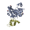
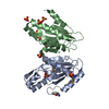
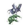
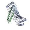
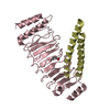
There are two heterodimers in the asymmetric unit. The biological heterodimer belongs to the pairs: (i) Chain A and Chain B, and (ii) Chain C and Chain D.
-
Components
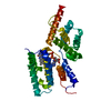
#1: Protein
E3ubiquitin-proteinligasesspH1 / Salmonella secreted protein H1 / Secreted effector protein sspH1
In the structure databanks used in Yorodumi, some data are registered as the other names, "COVID-19 virus" and "2019-nCoV". Here are the details of the virus and the list of structure data.
Jan 31, 2019. EMDB accession codes are about to change! (news from PDBe EMDB page)
EMDB accession codes are about to change! (news from PDBe EMDB page)
The allocation of 4 digits for EMDB accession codes will soon come to an end. Whilst these codes will remain in use, new EMDB accession codes will include an additional digit and will expand incrementally as the available range of codes is exhausted. The current 4-digit format prefixed with “EMD-” (i.e. EMD-XXXX) will advance to a 5-digit format (i.e. EMD-XXXXX), and so on. It is currently estimated that the 4-digit codes will be depleted around Spring 2019, at which point the 5-digit format will come into force.
The EM Navigator/Yorodumi systems omit the EMD- prefix.
Related info.:Q: What is EMD? / ID/Accession-code notation in Yorodumi/EM Navigator
Yorodumi is a browser for structure data from EMDB, PDB, SASBDB, etc.
This page is also the successor to EM Navigator detail page, and also detail information page/front-end page for Omokage search.
The word "yorodu" (or yorozu) is an old Japanese word meaning "ten thousand". "mi" (miru) is to see.
Related info.:EMDB / PDB / SASBDB / Comparison of 3 databanks / Yorodumi Search / Aug 31, 2016. New EM Navigator & Yorodumi / Yorodumi Papers / Jmol/JSmol / Function and homology information / Changes in new EM Navigator and Yorodumi
 Movie
Movie Controller

 Yorodumi
 Open data
 Basic information
 Components
 Keywords
 Function and homology information
 Salmonella enterica subsp. enterica serovar Typhimurium (bacteria)
 Homo sapiens (human)
 X-RAY DIFFRACTION /
  Authors
 Citation
 Structure visualization
 Downloads & links
 Other downloads


 PDBj
PDBj


 Assembly
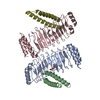

 Mass: 118.174 Da / Num. of mol.: 2 / Source method: obtained synthetically / Formula: C6H14O2
 Mass: 18.015 Da / Num. of mol.: 3 / Source method: isolated from a natural source / Formula: H2O
 Sample preparation
 / Beamline: 08ID-1 / Wavelength: 0.97949
  Processing