 Movie
Movie Controller


+ Open data

- Basic information
| Entry | Database: PDB / ID: 1nvq | ||||||
|---|---|---|---|---|---|---|---|
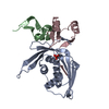
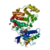
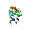
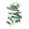
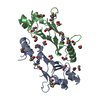
| Title | The Complex Structure Of Checkpoint Kinase Chk1/UCN-01 | ||||||
 Components Components |
| ||||||
 Keywords Keywords | TRANSFERASE / Chk1-UCN-01 COMPLEX | ||||||
| Function / homology |  Function and homology information Function and homology informationapoptotic process involved in development / negative regulation of G0 to G1 transition / histone H3T11 kinase activity / regulation of mitotic centrosome separation / negative regulation of mitotic nuclear division / mitotic G2/M transition checkpoint / inner cell mass cell proliferation / regulation of double-strand break repair via homologous recombination / nucleus organization / negative regulation of gene expression, epigenetic ...apoptotic process involved in development / negative regulation of G0 to G1 transition / histone H3T11 kinase activity / regulation of mitotic centrosome separation / negative regulation of mitotic nuclear division / mitotic G2/M transition checkpoint / inner cell mass cell proliferation / regulation of double-strand break repair via homologous recombination / nucleus organization / negative regulation of gene expression, epigenetic / peptidyl-threonine phosphorylation / Transcriptional Regulation by E2F6 / mitotic G2 DNA damage checkpoint signaling / Presynaptic phase of homologous DNA pairing and strand exchange / replicative senescence / Activation of ATR in response to replication stress / Chk1/Chk2(Cds1) mediated inactivation of Cyclin B:Cdk1 complex / signal transduction in response to DNA damage / positive regulation of cell cycle / DNA damage checkpoint signaling / regulation of signal transduction by p53 class mediator / replication fork / condensed nuclear chromosome / TP53 Regulates Transcription of DNA Repair Genes / cellular response to mechanical stimulus / Signaling by SCF-KIT / Ubiquitin-Mediated Degradation of Phosphorylated Cdc25A / G2/M DNA damage checkpoint / G2/M transition of mitotic cell cycle / regulation of cell population proliferation / Processing of DNA double-strand break ends / Regulation of TP53 Activity through Phosphorylation / DNA replication / protein phosphorylation / protein kinase activity / non-specific serine/threonine protein kinase / chromatin remodeling / protein domain specific binding / protein serine kinase activity / DNA repair / protein serine/threonine kinase activity / apoptotic process / DNA damage response / centrosome / chromatin / protein-containing complex / extracellular space / nucleoplasm / ATP binding / nucleus / cytoplasm / cytosol Similarity search - Function | ||||||
| Biological species |  Homo sapiens (human) Homo sapiens (human) | ||||||
| Method |  X-RAY DIFFRACTION / SYNCHROTRON / MOLECULAR REPLACEMENT / Resolution: 2 Å X-RAY DIFFRACTION / SYNCHROTRON / MOLECULAR REPLACEMENT / Resolution: 2 Å | ||||||
 Authors Authors | Zhao, B. / Bower, M.J. / McDevitt, P.J. / Zhao, H. / Davis, S.T. / Johanson, K.O. / Green, S.M. / Concha, N.O. / Zhou, B.B. | ||||||
 Citation Citation | Journal: J.Biol.Chem. / Year: 2002 Title: Structural Basis for Chk1 Inhibition by UCN-01 Authors: Zhao, B. / Bower, M.J. / McDevitt, P.J. / Zhao, H. / Davis, S.T. / Johanson, K.O. / Green, S.M. / Concha, N.O. / Zhou, B.B. | ||||||
| History |
| ||||||
| Remark 999 | SEQUENCE A five-residue peptide 301-305 was found with the enzyme. Residues were named based on the ...SEQUENCE A five-residue peptide 301-305 was found with the enzyme. Residues were named based on the electron density. The peptide could be the part of C-terminal missing residues. |
- Structure visualization
| Structure viewer | Molecule: MolmilJmol/JSmol |
|---|
- Downloads & links
-Download
| PDBx/mmCIF format | 1nvq.cif.gz | 72.8 KB | Display | PDBx/mmCIF format |
|---|---|---|---|---|
| PDB format | pdb1nvq.ent.gz | 52.7 KB | Display | PDB format |
| PDBx/mmJSON format | 1nvq.json.gz | Tree view | PDBx/mmJSON format | |
| Others |  Other downloads Other downloads |
-Validation report
| Arichive directory | https://data.pdbj.org/pub/pdb/validation_reports/nv/1nvqftp://data.pdbj.org/pub/pdb/validation_reports/nv/1nvq | HTTPS FTP |
|---|
-Related structure data
| Related structure data | 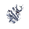 1nvrC 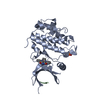 1nvsC 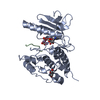 2phkS S: Starting model for refinement C: citing same article ( |
|---|---|
| Similar structure data |


-Links
 PDBj
PDBj


- Assembly
| Deposited unit | 
| ||||||||
|---|---|---|---|---|---|---|---|---|---|
| 1 |
| ||||||||
| Unit cell |
|
-Components
| #1: Protein | Mass: 33042.988 Da / Num. of mol.: 1 / Fragment: CHK1KD (RESIDUES 1-289) Source method: isolated from a genetically manipulated source Source: (gene. exp.) Homo sapiens (human) / Production host:  unidentified baculovirus / Strain (production host): sf9 unidentified baculovirus / Strain (production host): sf9References: UniProt: O14757, Transferases; Transferring phosphorus-containing groups; Phosphotransferases with an alcohol group as acceptor |
|---|---|
| #2: Protein/peptide | Mass: 433.457 Da / Num. of mol.: 1 / Source method: obtained synthetically |
| #3: Chemical | ChemComp-SO4 /   Mass: 96.063 Da / Num. of mol.: 1 / Source method: obtained synthetically / Formula: SO4 Mass: 96.063 Da / Num. of mol.: 1 / Source method: obtained synthetically / Formula: SO4 |
| #4: Chemical | ChemComp-UCN /   Mass: 482.530 Da / Num. of mol.: 1 / Source method: obtained synthetically / Formula: C28H26N4O4 Mass: 482.530 Da / Num. of mol.: 1 / Source method: obtained synthetically / Formula: C28H26N4O4 |
| #5: Water | ChemComp-HOH /  Mass: 18.015 Da / Num. of mol.: 192 / Source method: isolated from a natural source / Formula: H2O Mass: 18.015 Da / Num. of mol.: 192 / Source method: isolated from a natural source / Formula: H2O |
-Experimental details
-Experiment
| Experiment | Method: X-RAY DIFFRACTION / Number of used crystals: 1 |
|---|
- Sample preparation
| Crystal | Density Matthews: 2.54 Å3/Da / Density % sol: 51.12 % | ||||||||||||||||||||
|---|---|---|---|---|---|---|---|---|---|---|---|---|---|---|---|---|---|---|---|---|---|
| Crystal grow | Temperature: 293 K / Method: vapor diffusion, sitting drop / pH: 7.5 Details: PEG8000, ammonium sulfate, glycerol, pH 7.5, VAPOR DIFFUSION, SITTING DROP, temperature 293K | ||||||||||||||||||||
| Crystal grow | *PLUS Method: vapor diffusion | ||||||||||||||||||||
| Components of the solutions | *PLUS
|
-Data collection
| Diffraction | Mean temperature: 100 K |
|---|---|
| Diffraction source | Source: SYNCHROTRON / Site: APS  / Beamline: 17-ID / Wavelength: 1 / Beamline: 17-ID / Wavelength: 1 |
| Detector | Type: MARRESEARCH / Detector: CCD / Date: Mar 1, 2001 |
| Radiation | Protocol: SINGLE WAVELENGTH / Monochromatic (M) / Laue (L): M / Scattering type: x-ray |
| Radiation wavelength | Wavelength: 1 Å / Relative weight: 1 |
| Reflection | Resolution: 2→50 Å / Num. obs: 22177 / % possible obs: 97 % / Observed criterion σ(F): 1 / Observed criterion σ(I): 1 |
| Reflection | *PLUS Highest resolution: 2 Å / Num. obs: 22212 / % possible obs: 97 % / Redundancy: 3.4 % / Num. measured all: 75805 / Rmerge(I) obs: 0.065 |
- Processing
| Software |
| |||||||||||||||||||||||||
|---|---|---|---|---|---|---|---|---|---|---|---|---|---|---|---|---|---|---|---|---|---|---|---|---|---|---|
| Refinement | Method to determine structure: MOLECULAR REPLACEMENT Starting model: PDB ENTRY 2PHK Resolution: 2→20 Å / σ(F): 1 / Stereochemistry target values: Engh & Huber
| |||||||||||||||||||||||||
| Refinement step | Cycle: LAST / Resolution: 2→20 Å
| |||||||||||||||||||||||||
| Refine LS restraints |
| |||||||||||||||||||||||||
| Refinement | *PLUS Lowest resolution: 20 Å / % reflection Rfree: 6 % | |||||||||||||||||||||||||
| Solvent computation | *PLUS | |||||||||||||||||||||||||
| Displacement parameters | *PLUS | |||||||||||||||||||||||||
| Refine LS restraints | *PLUS Type: c_angle_deg / Dev ideal: 1.168 |
