 Movie
Movie Controller

[English] 日本語
 Yorodumi
- PDB-2phk: THE CRYSTAL STRUCTURE OF A PHOSPHORYLASE KINASE PEPTIDE SUBSTRATE... -
+ Open data

- Basic information
| Entry | Database: PDB / ID: 2phk | ||||||
|---|---|---|---|---|---|---|---|
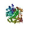
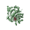
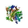
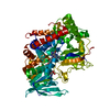
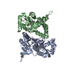
| Title | THE CRYSTAL STRUCTURE OF A PHOSPHORYLASE KINASE PEPTIDE SUBSTRATE COMPLEX: KINASE SUBSTRATE RECOGNITION | ||||||
 Components Components |
| ||||||
 Keywords Keywords | COMPLEX (TRANSFERASE/PEPTIDE) / CATALYTIC MECHANISM / DIMERIZATION / PHOSPHORYLASE KINASE / REVERSIBLE PHOSPHORYLISATION / SUBSTRATE RECOGNITION / COMPLEX (TRANSFERASE-PEPTIDE) / COMPLEX (TRANSFERASE-PEPTIDE) complex | ||||||
| Function / homology |  Function and homology information Function and homology informationphosphorylase kinase / phosphorylase kinase activity / phosphorylase kinase complex / tau-protein kinase / glycogen catabolic process / tau-protein kinase activity / skeletal muscle myofibril / calmodulin binding / non-specific serine/threonine protein kinase / protein serine kinase activity / ATP binding Similarity search - Function | ||||||
| Biological species |  | ||||||
| Method |  X-RAY DIFFRACTION / SYNCHROTRON / MOLECULAR REPLACEMENT / Resolution: 2.6 Å X-RAY DIFFRACTION / SYNCHROTRON / MOLECULAR REPLACEMENT / Resolution: 2.6 Å | ||||||
 Authors Authors | Lowe, E.D. / Noble, M.E.M. / Skamnaki, V.T. / Oikonomakos, N.G. / Owen, D.J. / Johnson, L.N. | ||||||
 Citation Citation | Journal: EMBO J. / Year: 1997 Title: The crystal structure of a phosphorylase kinase peptide substrate complex: kinase substrate recognition. Authors: Lowe, E.D. / Noble, M.E. / Skamnaki, V.T. / Oikonomakos, N.G. / Owen, D.J. / Johnson, L.N. #1: Journal: Structure / Year: 1995Title: Two Structures of the Catalytic Domain of Phosphorylase Kinase: An Active Protein Kinase Complexed with Substrate Analogue and Product Authors: Owen, D.J. / Noble, M.E. / Garman, E.F. / Papageorgiou, A.C. / Johnson, L.N. | ||||||
| History |
|
- Structure visualization
| Structure viewer | Molecule: MolmilJmol/JSmol |
|---|
- Downloads & links
-Download
| PDBx/mmCIF format | 2phk.cif.gz | 74.6 KB | Display | PDBx/mmCIF format |
|---|---|---|---|---|
| PDB format | pdb2phk.ent.gz | 54.5 KB | Display | PDB format |
| PDBx/mmJSON format | 2phk.json.gz | Tree view | PDBx/mmJSON format | |
| Others |  Other downloads Other downloads |
-Validation report
| Arichive directory | https://data.pdbj.org/pub/pdb/validation_reports/ph/2phkftp://data.pdbj.org/pub/pdb/validation_reports/ph/2phk | HTTPS FTP |
|---|
-Related structure data
| Related structure data | 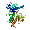 1phkS S: Starting model for refinement |
|---|---|
| Similar structure data |


-Links
 PDBj
PDBj- Assembly
| Deposited unit | 
| ||||||||
|---|---|---|---|---|---|---|---|---|---|
| 1 | 
| ||||||||
| Unit cell |
|
-Components
-Protein / Protein/peptide , 2 types, 2 molecules AB
| #1: Protein | Mass: 31983.758 Da / Num. of mol.: 1 / Fragment: CATALYTIC DOMAIN Source method: isolated from a genetically manipulated source Source: (gene. exp.)  |
|---|---|
| #2: Protein/peptide | Mass: 939.137 Da / Num. of mol.: 1 Source method: isolated from a genetically manipulated source |
-Non-polymers , 4 types, 90 molecules 


| #3: Chemical |  Mass: 54.938 Da / Num. of mol.: 2 / Source method: obtained synthetically / Formula: Mn Mass: 54.938 Da / Num. of mol.: 2 / Source method: obtained synthetically / Formula: Mn#4: Chemical | ChemComp-ATP / |  Mass: 507.181 Da / Num. of mol.: 1 / Source method: obtained synthetically / Formula: C10H16N5O13P3 / Comment: ATP, energy-carrying molecule*YM Mass: 507.181 Da / Num. of mol.: 1 / Source method: obtained synthetically / Formula: C10H16N5O13P3 / Comment: ATP, energy-carrying molecule*YM#5: Chemical | ChemComp-GOL / |  Mass: 92.094 Da / Num. of mol.: 1 / Source method: obtained synthetically / Formula: C3H8O3 Mass: 92.094 Da / Num. of mol.: 1 / Source method: obtained synthetically / Formula: C3H8O3#6: Water | ChemComp-HOH / | Mass: 18.015 Da / Num. of mol.: 86 / Source method: isolated from a natural source / Formula: H2O |
|---|
-Experimental details
-Experiment
| Experiment | Method: X-RAY DIFFRACTION / Number of used crystals: 1 |
|---|
- Sample preparation
| Crystal | Density Matthews: 2.7 Å3/Da / Density % sol: 54.4 % | ||||||||||||||||||||||||||||||||||||||||||||||||||||||||||||||||||||||||||||||||||||||||||||||||
|---|---|---|---|---|---|---|---|---|---|---|---|---|---|---|---|---|---|---|---|---|---|---|---|---|---|---|---|---|---|---|---|---|---|---|---|---|---|---|---|---|---|---|---|---|---|---|---|---|---|---|---|---|---|---|---|---|---|---|---|---|---|---|---|---|---|---|---|---|---|---|---|---|---|---|---|---|---|---|---|---|---|---|---|---|---|---|---|---|---|---|---|---|---|---|---|---|---|
| Crystal grow | pH: 6.9 / Details: pH 6.9 | ||||||||||||||||||||||||||||||||||||||||||||||||||||||||||||||||||||||||||||||||||||||||||||||||
| Crystal grow | *PLUS pH: 8.2 / Method: vapor diffusion, hanging drop | ||||||||||||||||||||||||||||||||||||||||||||||||||||||||||||||||||||||||||||||||||||||||||||||||
| Components of the solutions | *PLUS
|
-Data collection
| Diffraction | Mean temperature: 100 K |
|---|---|
| Diffraction source | Source: SYNCHROTRON / Site: ESRF  / Beamline: BM02 / Wavelength: 0.9 / Beamline: BM02 / Wavelength: 0.9 |
| Detector | Type: XRII / Detector: CCD AREA DETECTOR / Date: Mar 11, 1997 / Details: MIRRORS |
| Radiation | Monochromator: NI FILTER / Monochromatic (M) / Laue (L): M / Scattering type: x-ray |
| Radiation wavelength | Wavelength: 0.9 Å / Relative weight: 1 |
| Reflection | Resolution: 2.6→20 Å / Num. obs: 10062 / % possible obs: 86.3 % / Observed criterion σ(I): 2 / Redundancy: 2.15 % / Rmerge(I) obs: 0.098 / Rsym value: 0.089 / Net I/σ(I): 7.1 |
| Reflection shell | Resolution: 2.6→2.74 Å / Redundancy: 3.7 % / Rmerge(I) obs: 0.36 / Mean I/σ(I) obs: 1.3 / Rsym value: 0.397 / % possible all: 78.1 |
| Reflection | *PLUS Num. measured all: 21644 |
| Reflection shell | *PLUS % possible obs: 78.1 % |
- Processing
| Software |
| ||||||||||||||||
|---|---|---|---|---|---|---|---|---|---|---|---|---|---|---|---|---|---|
| Refinement | Method to determine structure: MOLECULAR REPLACEMENT Starting model: PDB ENTRY 1PHK Resolution: 2.6→25 Å / σ(F): 0.3
| ||||||||||||||||
| Refinement step | Cycle: LAST / Resolution: 2.6→25 Å
| ||||||||||||||||
| Software | *PLUS Name: REFMAC / Classification: refinement | ||||||||||||||||
| Refinement | *PLUS | ||||||||||||||||
| Solvent computation | *PLUS | ||||||||||||||||
| Displacement parameters | *PLUS | ||||||||||||||||
| Refine LS restraints | *PLUS
|
