 Movie
Movie Controller


+ Open data

- Basic information
| Entry | Database: PDB / ID: 5was | ||||||
|---|---|---|---|---|---|---|---|
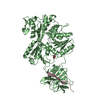
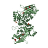
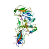
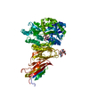
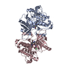
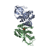
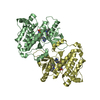
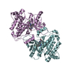
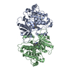
| Title | Corynebacterium glutamicum Hydrolyzed Homoserine kinase | ||||||
 Components Components | (Homoserine kinase) x 3 | ||||||
 Keywords Keywords | TRANSFERASE / Corynebacterium glutamicum / Homoserine Kinase / L-threonine / L-homoserine / Magnesium | ||||||
| Function / homology |  Function and homology information Function and homology informationhomoserine kinase / homoserine kinase activity / L-threonine biosynthetic process / ATP binding / cytoplasm Similarity search - Function | ||||||
| Biological species |  Corynebacterium glutamicum (bacteria) Corynebacterium glutamicum (bacteria) | ||||||
| Method |  X-RAY DIFFRACTION / SYNCHROTRON / MOLECULAR REPLACEMENT / Resolution: 1.799 Å X-RAY DIFFRACTION / SYNCHROTRON / MOLECULAR REPLACEMENT / Resolution: 1.799 Å | ||||||
 Authors Authors | Petit, C. / Ronning, D.R. | ||||||
 Citation Citation | Journal: ACS Omega / Year: 2018 Title: Reduction of Feedback Inhibition in Homoserine Kinase (ThrB) ofCorynebacterium glutamicumEnhances l-Threonine Biosynthesis. Authors: Petit, C. / Kim, Y. / Lee, S.K. / Brown, J. / Larsen, E. / Ronning, D.R. / Suh, J.W. / Kang, C.M. | ||||||
| History |
|
- Structure visualization
| Structure viewer | Molecule: MolmilJmol/JSmol |
|---|
- Downloads & links
-Download
| PDBx/mmCIF format | 5was.cif.gz | 69 KB | Display | PDBx/mmCIF format |
|---|---|---|---|---|
| PDB format | pdb5was.ent.gz | 48.5 KB | Display | PDB format |
| PDBx/mmJSON format | 5was.json.gz | Tree view | PDBx/mmJSON format | |
| Others |  Other downloads Other downloads |
-Validation report
| Arichive directory | https://data.pdbj.org/pub/pdb/validation_reports/wa/5wasftp://data.pdbj.org/pub/pdb/validation_reports/wa/5was | HTTPS FTP |
|---|
-Related structure data
| Related structure data | 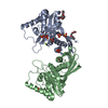 5watSC S: Starting model for refinement C: citing same article ( |
|---|---|
| Similar structure data |

-Links
 PDBj
PDBj- Assembly
| Deposited unit | 
| ||||||||
|---|---|---|---|---|---|---|---|---|---|
| 1 | 
| ||||||||
| Unit cell |
|
-Components
| #1: Protein | Mass: 18769.039 Da / Num. of mol.: 1 Source method: isolated from a genetically manipulated source Source: (gene. exp.) Corynebacterium glutamicum (bacteria) / Gene: thrB, Cgl1184, cg1338 / Production host: Escherichia coli BL21 / References: UniProt: P07128, homoserine kinase |
|---|---|
| #2: Protein | Mass: 6464.402 Da / Num. of mol.: 1 Source method: isolated from a genetically manipulated source Source: (gene. exp.) Corynebacterium glutamicum (bacteria) / Gene: thrB, Cgl1184, cg1338 / Production host: |
| #3: Protein | Mass: 8281.354 Da / Num. of mol.: 1 Source method: isolated from a genetically manipulated source Source: (gene. exp.) Corynebacterium glutamicum (bacteria) / Gene: thrB, Cgl1184, cg1338 / Production host: |
| #4: Chemical | ChemComp-PO4 /   Mass: 94.971 Da / Num. of mol.: 1 / Source method: obtained synthetically / Formula: PO4 Mass: 94.971 Da / Num. of mol.: 1 / Source method: obtained synthetically / Formula: PO4 |
| #5: Water | ChemComp-HOH /  Mass: 18.015 Da / Num. of mol.: 61 / Source method: isolated from a natural source / Formula: H2O Mass: 18.015 Da / Num. of mol.: 61 / Source method: isolated from a natural source / Formula: H2O |
-Experimental details
-Experiment
| Experiment | Method: X-RAY DIFFRACTION / Number of used crystals: 1 |
|---|
- Sample preparation
| Crystal | Density Matthews: 2.19 Å3/Da / Density % sol: 43.75 % |
|---|---|
| Crystal grow | Temperature: 293 K / Method: vapor diffusion, hanging drop / pH: 7.5 Details: The CglThrB protein concentrated to 16.2 mg/mL in 20 mM Tris pH 7.5, 150 mM NaCl, 50 mM KCl and 50 mM MgCl2 was used for crystallization experiments. Drops were equilibrated against a 100 ...Details: The CglThrB protein concentrated to 16.2 mg/mL in 20 mM Tris pH 7.5, 150 mM NaCl, 50 mM KCl and 50 mM MgCl2 was used for crystallization experiments. Drops were equilibrated against a 100 microLitter of well solution containing 0.25 M ammonium sulfate, 25 % PEG 3,350 and 0.1 M HEPES pH 7.5 |
-Data collection
| Diffraction | Mean temperature: 100 K |
|---|---|
| Diffraction source | Source: SYNCHROTRON / Site: APS  / Beamline: 21-ID-F / Wavelength: 0.97872 Å / Beamline: 21-ID-F / Wavelength: 0.97872 Å |
| Detector | Type: MARMOSAIC 325 mm CCD / Detector: CCD / Date: Oct 16, 2015 |
| Radiation | Protocol: SINGLE WAVELENGTH / Monochromatic (M) / Laue (L): M / Scattering type: x-ray |
| Radiation wavelength | Wavelength: 0.97872 Å / Relative weight: 1 |
| Reflection | Resolution: 1.799→50 Å / Num. obs: 27709 / % possible obs: 97.8 % / Redundancy: 23.4 % / Rmerge(I) obs: 0.061 / Net I/σ(I): 43.7 |
| Reflection shell | Highest resolution: 1.8 Å / Redundancy: 19.5 % / Rmerge(I) obs: 0.262 / Mean I/σ(I) obs: 6.3 / Num. unique obs: 2537 / % possible all: 92.8 |
- Processing
| Software |
| |||||||||||||||||||||||||||||||||||||||||||||||||||||||||||||||||||||||||||||||||||||||||||||||||||||||||
|---|---|---|---|---|---|---|---|---|---|---|---|---|---|---|---|---|---|---|---|---|---|---|---|---|---|---|---|---|---|---|---|---|---|---|---|---|---|---|---|---|---|---|---|---|---|---|---|---|---|---|---|---|---|---|---|---|---|---|---|---|---|---|---|---|---|---|---|---|---|---|---|---|---|---|---|---|---|---|---|---|---|---|---|---|---|---|---|---|---|---|---|---|---|---|---|---|---|---|---|---|---|---|---|---|---|---|
| Refinement | Method to determine structure: MOLECULAR REPLACEMENT Starting model: 5WAT Resolution: 1.799→34.967 Å / SU ML: 0.23 / Cross valid method: NONE / σ(F): 1.34 / Phase error: 34.36
| |||||||||||||||||||||||||||||||||||||||||||||||||||||||||||||||||||||||||||||||||||||||||||||||||||||||||
| Solvent computation | Shrinkage radii: 0.9 Å / VDW probe radii: 1.11 Å | |||||||||||||||||||||||||||||||||||||||||||||||||||||||||||||||||||||||||||||||||||||||||||||||||||||||||
| Refinement step | Cycle: LAST / Resolution: 1.799→34.967 Å
| |||||||||||||||||||||||||||||||||||||||||||||||||||||||||||||||||||||||||||||||||||||||||||||||||||||||||
| Refine LS restraints |
| |||||||||||||||||||||||||||||||||||||||||||||||||||||||||||||||||||||||||||||||||||||||||||||||||||||||||
| LS refinement shell |
|
