 Movie
Movie Controller

[English] 日本語
 Yorodumi
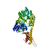
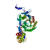
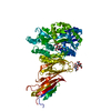
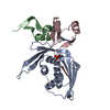
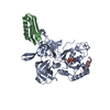
- PDB-2j42: low quality crystal structure of the transport component C2-II of... -
+ Open data

- Basic information
| Entry | Database: PDB / ID: 2j42 | ||||||
|---|---|---|---|---|---|---|---|
| Title | low quality crystal structure of the transport component C2-II of the C2-toxin from Clostridium botulinum | ||||||
 Components Components | C2 TOXIN COMPONENT-II | ||||||
 Keywords Keywords | TOXIN / CLOSTRIDIUM BOTULINUM / C2-II | ||||||
| Function / homology |  Function and homology information Function and homology information | ||||||
| Biological species |   CLOSTRIDIUM BOTULINUM (bacteria) CLOSTRIDIUM BOTULINUM (bacteria) | ||||||
| Method |  X-RAY DIFFRACTION / SYNCHROTRON / MOLECULAR REPLACEMENT / Resolution: 3.13 Å X-RAY DIFFRACTION / SYNCHROTRON / MOLECULAR REPLACEMENT / Resolution: 3.13 Å | ||||||
 Authors Authors | Schleberger, C. / Hochmann, H. / Barth, H. / Aktories, K. / Schulz, G.E. | ||||||
 Citation Citation | Journal: J.Mol.Biol. / Year: 2006 Title: Structure and Action of the Binary C2 Toxin from Clostridium Botulinum. Authors: Schleberger, C. / Hochmann, H. / Barth, H. / Aktories, K. / Schulz, G.E. | ||||||
| History |
| ||||||
| Remark 700 | SHEET THE SHEET STRUCTURE OF THIS MOLECULE IS BIFURCATED. IN ORDER TO REPRESENT THIS FEATURE IN ... SHEET THE SHEET STRUCTURE OF THIS MOLECULE IS BIFURCATED. IN ORDER TO REPRESENT THIS FEATURE IN THE SHEET RECORDS BELOW, TWO SHEETS ARE DEFINED. |
- Structure visualization
| Structure viewer | Molecule: MolmilJmol/JSmol |
|---|
- Downloads & links
-Download
| PDBx/mmCIF format | 2j42.cif.gz | 113.4 KB | Display | PDBx/mmCIF format |
|---|---|---|---|---|
| PDB format | pdb2j42.ent.gz | 85.2 KB | Display | PDB format |
| PDBx/mmJSON format | 2j42.json.gz | Tree view | PDBx/mmJSON format | |
| Others |  Other downloads Other downloads |
-Validation report
| Arichive directory | https://data.pdbj.org/pub/pdb/validation_reports/j4/2j42ftp://data.pdbj.org/pub/pdb/validation_reports/j4/2j42 | HTTPS FTP |
|---|
-Related structure data
| Related structure data | 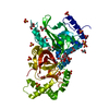 2j3vC  2j3xC  2j3zC  1accS S: Starting model for refinement C: citing same article ( |
|---|---|
| Similar structure data |


-Links
 PDBj
PDBj
- Assembly
| Deposited unit | 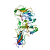
| ||||||||
|---|---|---|---|---|---|---|---|---|---|
| 1 |
| ||||||||
| Unit cell |
|
-Components
| #1: Protein | Mass: 80766.562 Da / Num. of mol.: 1 Source method: isolated from a genetically manipulated source Source: (gene. exp.) CLOSTRIDIUM BOTULINUM (bacteria) / Plasmid: PGEX-2T / Production host: |
|---|
-Experimental details
-Experiment
| Experiment | Method: X-RAY DIFFRACTION / Number of used crystals: 1 |
|---|
- Sample preparation
| Crystal | Density Matthews: 2.6 Å3/Da / Density % sol: 52 % |
|---|---|
| Crystal grow | pH: 4.6 Details: PROTEIN: 3 MG/ML IN H2O RESERVOIR: 0.1 M SODIUM ACETATE PH 4.6, 1.5 M (NH4)2SO4, 5 % ISOPROPANOL, 3 % ETHANOL |
-Data collection
| Diffraction | Mean temperature: 100 K |
|---|---|
| Diffraction source | Source: SYNCHROTRON / Site: SLS  / Beamline: X06SA / Wavelength: 0.9795 / Beamline: X06SA / Wavelength: 0.9795 |
| Detector | Type: MARRESEARCH / Detector: CCD |
| Radiation | Protocol: SINGLE WAVELENGTH / Monochromatic (M) / Laue (L): M / Scattering type: x-ray |
| Radiation wavelength | Wavelength: 0.9795 Å / Relative weight: 1 |
| Reflection | Resolution: 3.1→40 Å / Num. obs: 15486 / % possible obs: 99.6 % / Observed criterion σ(I): -3 / Redundancy: 9.5 % / Rmerge(I) obs: 0.08 / Net I/σ(I): 21.5 |
| Reflection shell | Resolution: 3.13→3.22 Å / Rmerge(I) obs: 0.44 / Mean I/σ(I) obs: 5.7 / % possible all: 100 |
- Processing
| Software |
| ||||||||||||||||||||
|---|---|---|---|---|---|---|---|---|---|---|---|---|---|---|---|---|---|---|---|---|---|
| Refinement | Method to determine structure: MOLECULAR REPLACEMENT Starting model: PDB ENTRY 1ACC Resolution: 3.13→30 Å / Cross valid method: THROUGHOUT / σ(F): 0 Details: INCOMPLETE MODEL, MISSING ATOM STRUCTURE WAS USED. MODEL IS OF LOW QUALITY DUE TO THE LOW RESOLUTION, NO FURTHER REFINEMENT WAS POSSIBLE.
| ||||||||||||||||||||
| Refinement step | Cycle: LAST / Resolution: 3.13→30 Å
|
