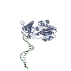
 Movie
Movie Controller

[English] 日本語
 Yorodumi
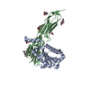
- PDB-2qn6: Structure of an archaeal heterotrimeric initiation factor 2 revea... -
+ Open data

- Basic information
| Entry | Database: PDB / ID: 2qn6 | ||||||
|---|---|---|---|---|---|---|---|
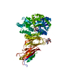
| Title | Structure of an archaeal heterotrimeric initiation factor 2 reveals a nucleotide state between the GTP and the GDP states | ||||||
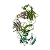
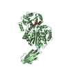
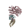
 Components Components | (Translation initiation factor 2 ...) x 3 | ||||||
 Keywords Keywords | TRANSLATION / initiation of translation / GTP-binding / Initiation factor / Nucleotide-binding / Protein biosynthesis / RNA-binding | ||||||
| Function / homology |  Function and homology information Function and homology informationformation of translation preinitiation complex / protein-synthesizing GTPase / translation elongation factor activity / translation initiation factor activity / translational initiation / ribosome binding / tRNA binding / GTPase activity / GTP binding / RNA binding / metal ion binding Similarity search - Function | ||||||
| Biological species |   Sulfolobus solfataricus (archaea) Sulfolobus solfataricus (archaea) | ||||||
| Method |  X-RAY DIFFRACTION / SYNCHROTRON / MOLECULAR REPLACEMENT / Resolution: 2.15 Å X-RAY DIFFRACTION / SYNCHROTRON / MOLECULAR REPLACEMENT / Resolution: 2.15 Å | ||||||
 Authors Authors | Mechulam, Y. / Yatime, L. / Blanquet, S. / Schmitt, E. | ||||||
 Citation Citation | Journal: Proc.Natl.Acad.Sci.Usa / Year: 2007 Title: Structure of an archaeal heterotrimeric initiation factor 2 reveals a nucleotide state between the GTP and the GDP states. Authors: Yatime, L. / Mechulam, Y. / Blanquet, S. / Schmitt, E. | ||||||
| History |
|
- Structure visualization
| Structure viewer | Molecule: MolmilJmol/JSmol |
|---|
- Downloads & links
-Download
| PDBx/mmCIF format | 2qn6.cif.gz | 118.6 KB | Display | PDBx/mmCIF format |
|---|---|---|---|---|
| PDB format | pdb2qn6.ent.gz | 89 KB | Display | PDB format |
| PDBx/mmJSON format | 2qn6.json.gz | Tree view | PDBx/mmJSON format | |
| Others |  Other downloads Other downloads |
-Validation report
| Arichive directory | https://data.pdbj.org/pub/pdb/validation_reports/qn/2qn6ftp://data.pdbj.org/pub/pdb/validation_reports/qn/2qn6 | HTTPS FTP |
|---|
-Related structure data
| Related structure data |  2qmuC  2ahoS S: Starting model for refinement C: citing same article ( |
|---|---|
| Similar structure data |


-Links
 PDBj
PDBj


- Assembly
| Deposited unit | 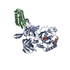
| ||||||||
|---|---|---|---|---|---|---|---|---|---|
| 1 |
| ||||||||
| Unit cell |
|
-Components
-Translation initiation factor 2 ... , 3 types, 3 molecules ABC
| #1: Protein | Mass: 45718.035 Da / Num. of mol.: 1 Source method: isolated from a genetically manipulated source Source: (gene. exp.) Sulfolobus solfataricus (archaea) / Strain: P2 / Gene: eif2g / Plasmid: pET3alpa / Production host:  |
|---|---|
| #2: Protein | Mass: 10440.162 Da / Num. of mol.: 1 / Fragment: domain 3, res 175 to 265 Source method: isolated from a genetically manipulated source Source: (gene. exp.) Sulfolobus solfataricus (archaea) / Strain: P2 / Gene: eif2a / Plasmid: pET3alpa / Production host: |
| #3: Protein/peptide | Mass: 2190.494 Da / Num. of mol.: 1 / Fragment: helix 1, res 2 to 19 Source method: isolated from a genetically manipulated source Source: (gene. exp.) Sulfolobus solfataricus (archaea) / Strain: P2 / Gene: eif2b / Plasmid: pET3alpa / Production host: |
-Non-polymers , 3 types, 255 molecules 


| #4: Chemical | ChemComp-MG /  Mass: 24.305 Da / Num. of mol.: 1 / Source method: obtained synthetically / Formula: Mg Mass: 24.305 Da / Num. of mol.: 1 / Source method: obtained synthetically / Formula: Mg |
|---|---|
| #5: Chemical | ChemComp-GDP /  Type: RNA linking / Mass: 443.201 Da / Num. of mol.: 1 / Source method: obtained synthetically / Formula: C10H15N5O11P2 / Comment: GDP, energy-carrying molecule*YM Type: RNA linking / Mass: 443.201 Da / Num. of mol.: 1 / Source method: obtained synthetically / Formula: C10H15N5O11P2 / Comment: GDP, energy-carrying molecule*YM |
| #6: Water | ChemComp-HOH / Mass: 18.015 Da / Num. of mol.: 253 / Source method: isolated from a natural source / Formula: H2O |
-Details
| Has protein modification | Y |
|---|
-Experimental details
-Experiment
| Experiment | Method: X-RAY DIFFRACTION / Number of used crystals: 1 |
|---|
- Sample preparation
| Crystal | Density Matthews: 2.66 Å3/Da / Density % sol: 53.72 % |
|---|---|
| Crystal grow | Temperature: 297 K / Method: vapor diffusion, sitting drop / pH: 7.5 Details: 16 % PEG 3350; 0.1 M Magnesium formate, pH 7.5, VAPOR DIFFUSION, SITTING DROP, temperature 297K |
-Data collection
| Diffraction | Mean temperature: 100 K |
|---|---|
| Diffraction source | Source: SYNCHROTRON / Site: ESRF  / Beamline: ID14-4 / Wavelength: 0.9795 Å / Beamline: ID14-4 / Wavelength: 0.9795 Å |
| Detector | Type: ADSC QUANTUM 315 / Detector: CCD / Date: Sep 6, 2006 |
| Radiation | Monochromator: DIAMOND / Protocol: SINGLE WAVELENGTH / Monochromatic (M) / Laue (L): M / Scattering type: x-ray |
| Radiation wavelength | Wavelength: 0.9795 Å / Relative weight: 1 |
| Reflection | Resolution: 2.15→100 Å / Num. all: 31835 / Num. obs: 31835 / % possible obs: 95.3 % / Redundancy: 2.3 % / Biso Wilson estimate: 39.6 Å2 / Rsym value: 0.052 / Net I/σ(I): 7.4 |
| Reflection shell | Resolution: 2.15→2.27 Å / Redundancy: 1.9 % / Mean I/σ(I) obs: 2.2 / Num. unique all: 4678 / Rsym value: 0.32 / % possible all: 96.6 |
- Processing
| Software |
| |||||||||||||||||||||||||
|---|---|---|---|---|---|---|---|---|---|---|---|---|---|---|---|---|---|---|---|---|---|---|---|---|---|---|
| Refinement | Method to determine structure: MOLECULAR REPLACEMENT Starting model: chain A from 2aho Resolution: 2.15→100 Å / Isotropic thermal model: isotropic / Cross valid method: THROUGHOUT / σ(F): 2 / Stereochemistry target values: Engh & Huber
| |||||||||||||||||||||||||
| Displacement parameters | Biso mean: 46 Å2
| |||||||||||||||||||||||||
| Refinement step | Cycle: LAST / Resolution: 2.15→100 Å
| |||||||||||||||||||||||||
| Refine LS restraints |
| |||||||||||||||||||||||||
| LS refinement shell | Resolution: 2.15→2.17 Å
|
