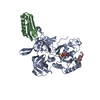
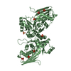
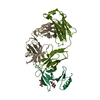
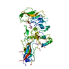
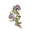
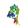
Maltodextrin-binding protein,Maltodextrin-binding protein,Maltodextrin-binding protein,ADAMTS13 CUB12,A disintegrin and metalloproteinase with thrombospondin motifs 13,A disintegrin and metalloproteinase with thrombospondin motifs 13,A disintegrin and metalloproteinase with thrombospondin motifs 13
Keywords
HYDROLASE / ADAMTS13 / CUB / Metalloprotease
Function / homology
Function and homology information
ADAMTS13 endopeptidase / Defective B3GALTL causes PpS / O-glycosylation of TSR domain-containing proteins / glycoprotein metabolic process / Enhanced cleavage of VWF variant by ADAMTS13 / Defective VWF cleavage by ADAMTS13 variant / response to potassium ion / Platelet Adhesion to exposed collagen / response to amine / peptide catabolic process ...ADAMTS13 endopeptidase / Defective B3GALTL causes PpS / O-glycosylation of TSR domain-containing proteins / glycoprotein metabolic process / Enhanced cleavage of VWF variant by ADAMTS13 / Defective VWF cleavage by ADAMTS13 variant / response to potassium ion / Platelet Adhesion to exposed collagen / response to amine / peptide catabolic process / carbohydrate transmembrane transporter activity / extracellular matrix organization / cellular response to interleukin-4 / cell-matrix adhesion / protein catabolic process / integrin-mediated signaling pathway / protein processing / metalloendopeptidase activity / platelet activation / cellular response to type II interferon / integrin binding / cellular response to tumor necrosis factor / response to toxic substance / metallopeptidase activity / blood coagulation / outer membrane-bounded periplasmic space / extracellular matrix / cellular response to lipopolysaccharide / endoplasmic reticulum lumen / calcium ion binding / cell surface / proteolysis / : / extracellular region / zinc ion binding Similarity search - Function
A: Maltodextrin-binding protein,Maltodextrin-binding protein,Maltodextrin-binding protein,ADAMTS13 CUB12,A disintegrin and metalloproteinase with thrombospondin motifs 13,A disintegrin and metalloproteinase with thrombospondin motifs 13,A disintegrin and metalloproteinase with thrombospondin motifs 13 hetero molecules
Resolution: 2.8→70 Å / Cor.coef. Fo:Fc: 0.917 / Cor.coef. Fo:Fc free: 0.807 / SU B: 24.339 / SU ML: 0.436 / Cross valid method: THROUGHOUT / σ(F): 0 / ESU R Free: 0.627 / Stereochemistry target values: MAXIMUM LIKELIHOOD Details: HYDROGENS HAVE BEEN ADDED IN THE RIDING POSITIONS U VALUES : REFINED INDIVIDUALLY
Rfactor
Num. reflection
% reflection
Selection details
Rfree
0.2917
566
4.7 %
RANDOM
Rwork
0.201
-
-
-
obs
0.2053
11438
58.7 %
-
Solvent computation
Ion probe radii: 0.8 Å / Shrinkage radii: 0.8 Å / VDW probe radii: 1.2 Å / Solvent model: MASK
In the structure databanks used in Yorodumi, some data are registered as the other names, "COVID-19 virus" and "2019-nCoV". Here are the details of the virus and the list of structure data.
Jan 31, 2019. EMDB accession codes are about to change! (news from PDBe EMDB page)
EMDB accession codes are about to change! (news from PDBe EMDB page)
The allocation of 4 digits for EMDB accession codes will soon come to an end. Whilst these codes will remain in use, new EMDB accession codes will include an additional digit and will expand incrementally as the available range of codes is exhausted. The current 4-digit format prefixed with “EMD-” (i.e. EMD-XXXX) will advance to a 5-digit format (i.e. EMD-XXXXX), and so on. It is currently estimated that the 4-digit codes will be depleted around Spring 2019, at which point the 5-digit format will come into force.
The EM Navigator/Yorodumi systems omit the EMD- prefix.
Related info.:Q: What is EMD? / ID/Accession-code notation in Yorodumi/EM Navigator
Yorodumi is a browser for structure data from EMDB, PDB, SASBDB, etc.
This page is also the successor to EM Navigator detail page, and also detail information page/front-end page for Omokage search.
The word "yorodu" (or yorozu) is an old Japanese word meaning "ten thousand". "mi" (miru) is to see.
Related info.:EMDB / PDB / SASBDB / Comparison of 3 databanks / Yorodumi Search / Aug 31, 2016. New EM Navigator & Yorodumi / Yorodumi Papers / Jmol/JSmol / Function and homology information / Changes in new EM Navigator and Yorodumi
 Movie
Movie Controller


 Open data
 Basic information
 Components
 Keywords
 Function and homology information
 Homo sapiens (human)
 X-RAY DIFFRACTION /
  Authors
 United Kingdom, 1items
  Citation
 Structure visualization
 Downloads & links
 Other downloads
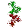


 PDBj
PDBj


 Assembly
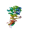

 Mass: 18.015 Da / Num. of mol.: 26 / Source method: isolated from a natural source / Formula: H2O
 Sample preparation
 Processing