 Movie
Movie Controller

[English] 日本語
 Yorodumi
- PDB-2fvq: A Structural Study of the CA Dinucleotide Step in the Integrase P... -
+ Open data

- Basic information
| Entry | Database: PDB / ID: 2fvq | ||||||
|---|---|---|---|---|---|---|---|
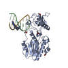
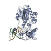
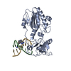
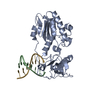
| Title | A Structural Study of the CA Dinucleotide Step in the Integrase Processing Site of Moloney Murine Leukemia Virus | ||||||
 Components Components |
| ||||||
 Keywords Keywords | TRANSFERASE/DNA / LTR / MMLV / TRANSFERASE-DNA COMPLEX | ||||||
| Function / homology |  Function and homology information Function and homology informationretroviral 3' processing activity / host cell late endosome membrane / DNA catabolic process / Hydrolases; Acting on peptide bonds (peptidases); Aspartic endopeptidases / ribonuclease H / virion assembly / protein-DNA complex / viral genome integration into host DNA / establishment of integrated proviral latency / RNA-directed DNA polymerase ...retroviral 3' processing activity / host cell late endosome membrane / DNA catabolic process / Hydrolases; Acting on peptide bonds (peptidases); Aspartic endopeptidases / ribonuclease H / virion assembly / protein-DNA complex / viral genome integration into host DNA / establishment of integrated proviral latency / RNA-directed DNA polymerase / host multivesicular body / RNA-directed DNA polymerase activity / RNA-DNA hybrid ribonuclease activity / Transferases; Transferring phosphorus-containing groups; Nucleotidyltransferases / viral nucleocapsid / DNA recombination / DNA-directed DNA polymerase / structural constituent of virion / aspartic-type endopeptidase activity / Hydrolases; Acting on ester bonds / DNA-directed DNA polymerase activity / symbiont-mediated suppression of host gene expression / symbiont entry into host cell / host cell plasma membrane / proteolysis / DNA binding / RNA binding / zinc ion binding Similarity search - Function | ||||||
| Biological species |  Moloney murine leukemia virus Moloney murine leukemia virus | ||||||
| Method |  X-RAY DIFFRACTION / MOLECULAR REPLACEMENT / Resolution: 2.3 Å X-RAY DIFFRACTION / MOLECULAR REPLACEMENT / Resolution: 2.3 Å | ||||||
 Authors Authors | Montano, S.P. / Cote, M.L. / Roth, M.J. / Georgiadis, M.M. | ||||||
 Citation Citation | Journal: Nucleic Acids Res. / Year: 2006 Title: Crystal structures of oligonucleotides including the integrase processing site of the Moloney murine leukemia virus. Authors: Montano, S.P. / Cote, M.L. / Roth, M.J. / Georgiadis, M.M. | ||||||
| History |
|
- Structure visualization
| Structure viewer | Molecule: MolmilJmol/JSmol |
|---|
- Downloads & links
-Download
| PDBx/mmCIF format | 2fvq.cif.gz | 84.2 KB | Display | PDBx/mmCIF format |
|---|---|---|---|---|
| PDB format | pdb2fvq.ent.gz | 60.5 KB | Display | PDB format |
| PDBx/mmJSON format | 2fvq.json.gz | Tree view | PDBx/mmJSON format | |
| Others |  Other downloads Other downloads |
-Validation report
| Arichive directory | https://data.pdbj.org/pub/pdb/validation_reports/fv/2fvqftp://data.pdbj.org/pub/pdb/validation_reports/fv/2fvq | HTTPS FTP |
|---|
-Related structure data
| Related structure data | 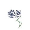 2fvpC  2fvrC 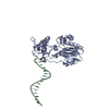 2fvsC 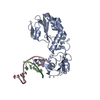 1d1uS C: citing same article ( S: Starting model for refinement |
|---|---|
| Similar structure data |


-Links
 PDBj
PDBj


- Assembly
| Deposited unit | 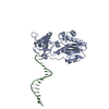
| ||||||||
|---|---|---|---|---|---|---|---|---|---|
| 1 | 
| ||||||||
| Unit cell |
| ||||||||
| Details | The second part of the biological assembly is generated by performing this transformation on the asymmetric unit: symmetry operator: -x, -y, z translation vector: 2 1 -1 |
-Components
| #1: DNA chain | Mass: 4896.216 Da / Num. of mol.: 1 / Source method: obtained synthetically |
|---|---|
| #2: Protein | Mass: 28934.287 Da / Num. of mol.: 1 Source method: isolated from a genetically manipulated source Source: (gene. exp.) Moloney murine leukemia virus / Genus: Gammaretrovirus / Species: Murine leukemia virus / Gene: pol / Plasmid: pET15b / Production host:  |
| #3: Water | ChemComp-HOH /  Mass: 18.015 Da / Num. of mol.: 130 / Source method: isolated from a natural source / Formula: H2O Mass: 18.015 Da / Num. of mol.: 130 / Source method: isolated from a natural source / Formula: H2O |
-Experimental details
-Experiment
| Experiment | Method: X-RAY DIFFRACTION / Number of used crystals: 1 |
|---|
- Sample preparation
| Crystal | Density Matthews: 2.77 Å3/Da / Density % sol: 55.52 % | ||||||||||||||||||||||||||||||||||||
|---|---|---|---|---|---|---|---|---|---|---|---|---|---|---|---|---|---|---|---|---|---|---|---|---|---|---|---|---|---|---|---|---|---|---|---|---|---|
| Crystal grow | Temperature: 293 K / Method: vapor diffusion, hanging drop / pH: 6.5 Details: PEG 4000, magnesium acetate, ADA, pH 6.5, VAPOR DIFFUSION, HANGING DROP, temperature 293K | ||||||||||||||||||||||||||||||||||||
| Components of the solutions |
|
-Data collection
| Diffraction | Mean temperature: 93 K |
|---|---|
| Diffraction source | Source: ROTATING ANODE / Type: RIGAKU / Wavelength: 1.54 Å |
| Detector | Type: RIGAKU RAXIS IV / Detector: IMAGE PLATE / Date: Apr 30, 2003 |
| Radiation | Monochromator: CONFOCAL MIRRORS / Protocol: SINGLE WAVELENGTH / Monochromatic (M) / Laue (L): M / Scattering type: x-ray |
| Radiation wavelength | Wavelength: 1.54 Å / Relative weight: 1 |
| Reflection | Resolution: 2.3→50 Å / Num. all: 17513 / Num. obs: 17275 / % possible obs: 98.7 % / Observed criterion σ(F): -3 / Observed criterion σ(I): -3 |
| Reflection shell | Resolution: 2.3→2.38 Å / % possible all: 99.6 |
- Processing
| Software |
| ||||||||||||||||||||
|---|---|---|---|---|---|---|---|---|---|---|---|---|---|---|---|---|---|---|---|---|---|
| Refinement | Method to determine structure: MOLECULAR REPLACEMENT Starting model: pdb entry 1d1u Resolution: 2.3→50 Å / σ(F): 0 / Stereochemistry target values: Engh & Huber
| ||||||||||||||||||||
| Refinement step | Cycle: LAST / Resolution: 2.3→50 Å
| ||||||||||||||||||||
| Refine LS restraints |
|
