 Movie
Movie Controller

[English] 日本語
 Yorodumi
- PDB-1n2d: Ternary complex of MLC1P bound to IQ2 and IQ3 of Myo2p, a class V... -
+ Open data

- Basic information
| Entry | Database: PDB / ID: 1n2d | ||||||
|---|---|---|---|---|---|---|---|
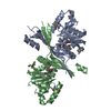
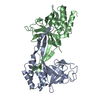
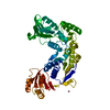
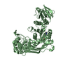
| Title | Ternary complex of MLC1P bound to IQ2 and IQ3 of Myo2p, a class V myosin | ||||||
 Components Components |
| ||||||
 Keywords Keywords | CELL CYCLE / PROTEIN-PEPTIDE COMPLEX / IQ MOTIF / MYOSIN LIGHT CHAIN | ||||||
| Function / homology |  Function and homology information Function and homology informationprotein localization to cell division site involved in mitotic actomyosin contractile ring assembly / MIH complex / RHO GTPases activate PAKs / regulation of actomyosin contractile ring contraction / peroxisome inheritance / regulation of cell wall organization or biogenesis / myosin II heavy chain binding / RHOT1 GTPase cycle / Myo2p-Vac17p-Vac8p transport complex / RHOT2 GTPase cycle ...protein localization to cell division site involved in mitotic actomyosin contractile ring assembly / MIH complex / RHO GTPases activate PAKs / regulation of actomyosin contractile ring contraction / peroxisome inheritance / regulation of cell wall organization or biogenesis / myosin II heavy chain binding / RHOT1 GTPase cycle / Myo2p-Vac17p-Vac8p transport complex / RHOT2 GTPase cycle / membrane addition at site of cytokinesis / mitochondrion inheritance / site of polarized growth / mitotic actomyosin contractile ring assembly / RHOU GTPase cycle / meiotic nuclear membrane microtubule tethering complex / cellular bud neck contractile ring / myosin V complex / vesicle targeting / vacuole inheritance / vesicle transport along actin filament / incipient cellular bud site / cellular bud tip / septum digestion after cytokinesis / myosin V binding / Golgi inheritance / cellular bud neck / mating projection tip / fungal-type vacuole membrane / myosin II complex / vesicle docking involved in exocytosis / microfilament motor activity / actin filament bundle / filamentous actin / intracellular distribution of mitochondria / establishment of mitotic spindle orientation / transport vesicle / vesicle-mediated transport / actin filament organization / regulation of cytokinesis / small GTPase binding / actin filament binding / actin cytoskeleton / protein transport / vesicle / calmodulin binding / calcium ion binding / ATP binding / identical protein binding / membrane / plasma membrane / cytoplasm Similarity search - Function | ||||||
| Biological species |  | ||||||
| Method |  X-RAY DIFFRACTION / SYNCHROTRON / MAD / Resolution: 2 Å X-RAY DIFFRACTION / SYNCHROTRON / MAD / Resolution: 2 Å | ||||||
 Authors Authors | Terrak, M. / Wu, G. / Stafford, W.F. / Lu, R.C. / Dominguez, R. | ||||||
 Citation Citation | Journal: Proc.Natl.Acad.Sci.USA / Year: 2005 Title: Structure of the light chain-binding domain of myosin V. Authors: Terrak, M. / Rebowski, G. / Lu, R.C. / Grabarek, Z. / Dominguez, R. #1: Journal: Acta Crystallogr.,Sect.D / Year: 2002Title: Crystallisation, X-ray Characterixation and Selenomethionine Phasing of Mlc1p Bound to IQ motifs from Myosin V Authors: Terrak, M. / Otterbein, L.R. / Wu, W. / Palecanda, L.A. / Lu, R.C. / Dominguez, R. | ||||||
| History |
|
- Structure visualization
| Structure viewer | Molecule: MolmilJmol/JSmol |
|---|
- Downloads & links
-Download
| PDBx/mmCIF format | 1n2d.cif.gz | 84.2 KB | Display | PDBx/mmCIF format |
|---|---|---|---|---|
| PDB format | pdb1n2d.ent.gz | 64.6 KB | Display | PDB format |
| PDBx/mmJSON format | 1n2d.json.gz | Tree view | PDBx/mmJSON format | |
| Others |  Other downloads Other downloads |
-Validation report
| Arichive directory | https://data.pdbj.org/pub/pdb/validation_reports/n2/1n2dftp://data.pdbj.org/pub/pdb/validation_reports/n2/1n2d | HTTPS FTP |
|---|
-Related structure data
| Related structure data | |
|---|---|
| Similar structure data |


-Links
 PDBj
PDBj


- Assembly
| Deposited unit | 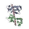
| ||||||||
|---|---|---|---|---|---|---|---|---|---|
| 1 |
| ||||||||
| Unit cell |
|
-Components
| #1: Protein | Mass: 16332.213 Da / Num. of mol.: 2 Source method: isolated from a genetically manipulated source Source: (gene. exp.) Gene: MLC1 / Plasmid: pAED4 / Species (production host): Escherichia coli / Production host:  #2: Protein/peptide | | Mass: 5543.434 Da / Num. of mol.: 1 / Source method: obtained synthetically Details: The peptide corresponding to IQ2 and IQ3 was chemically synthesized. THE sequence of the peptide is naturally found in saccharomyces cerevisiae (baker's yeast) MYO2P, a class V myosin. References: UniProt: P19524 #3: Water | ChemComp-HOH / |  Mass: 18.015 Da / Num. of mol.: 256 / Source method: isolated from a natural source / Formula: H2O Mass: 18.015 Da / Num. of mol.: 256 / Source method: isolated from a natural source / Formula: H2O |
|---|
-Experimental details
-Experiment
| Experiment | Method: X-RAY DIFFRACTION / Number of used crystals: 2 |
|---|
- Sample preparation
| Crystal | Density Matthews: 2.45 Å3/Da / Density % sol: 49.75 % | |||||||||||||||||||||||||||||||||||||||||||||||||
|---|---|---|---|---|---|---|---|---|---|---|---|---|---|---|---|---|---|---|---|---|---|---|---|---|---|---|---|---|---|---|---|---|---|---|---|---|---|---|---|---|---|---|---|---|---|---|---|---|---|---|
| Crystal grow | Temperature: 293 K / Method: vapor diffusion, hanging drop Details: PEG 5000 monomethyl ether, potassium fluoride, VAPOR DIFFUSION, HANGING DROP, temperature 293K | |||||||||||||||||||||||||||||||||||||||||||||||||
| Crystal grow | *PLUS Method: vapor diffusionDetails: Terrak, M., (2002) Acta Crystallogr.,Sect.D, 58, 1882. | |||||||||||||||||||||||||||||||||||||||||||||||||
| Components of the solutions | *PLUS
|
-Data collection
| Diffraction | Mean temperature: 100 K |
|---|---|
| Diffraction source | Source: SYNCHROTRON / Site: APS  / Beamline: 14-BM-C / Wavelength: 1 Å / Beamline: 14-BM-C / Wavelength: 1 Å |
| Detector | Type: ADSC QUANTUM 4 / Detector: CCD / Date: Aug 1, 2001 / Details: Bent conical Si-mirror (Rh coating) |
| Radiation | Monochromator: Bent Ge(111) monochromator / Protocol: SINGLE WAVELENGTH / Monochromatic (M) / Laue (L): M / Scattering type: x-ray |
| Radiation wavelength | Wavelength: 1 Å / Relative weight: 1 |
| Reflection | Resolution: 2→41.27 Å / Num. all: 25861 / Num. obs: 25861 / % possible obs: 99.5 % / Observed criterion σ(F): 0 / Observed criterion σ(I): -3 / Redundancy: 63.8 % / Biso Wilson estimate: 27.4 Å2 / Rmerge(I) obs: 0.098 / Net I/σ(I): 17 |
| Reflection shell | Resolution: 2→2.01 Å / Rmerge(I) obs: 0.423 / Mean I/σ(I) obs: 5 / % possible all: 99.1 |
- Processing
| Software |
| ||||||||||||||||||||||||||||||||||||||||||||||||||||||||||||
|---|---|---|---|---|---|---|---|---|---|---|---|---|---|---|---|---|---|---|---|---|---|---|---|---|---|---|---|---|---|---|---|---|---|---|---|---|---|---|---|---|---|---|---|---|---|---|---|---|---|---|---|---|---|---|---|---|---|---|---|---|---|
| Refinement | Method to determine structure: MAD / Resolution: 2→41.27 Å / Rfactor Rfree error: 0.007 / Data cutoff high absF: 1510444.66 / Data cutoff high rms absF: 1510444.66 / Data cutoff low absF: 0 / Isotropic thermal model: RESTRAINED / Cross valid method: THROUGHOUT / σ(F): 0 / Stereochemistry target values: Engh & Huber
| ||||||||||||||||||||||||||||||||||||||||||||||||||||||||||||
| Solvent computation | Solvent model: FLAT MODEL / Bsol: 56.5369 Å2 / ksol: 0.36759 e/Å3 | ||||||||||||||||||||||||||||||||||||||||||||||||||||||||||||
| Displacement parameters | Biso mean: 46.8 Å2
| ||||||||||||||||||||||||||||||||||||||||||||||||||||||||||||
| Refine analyze |
| ||||||||||||||||||||||||||||||||||||||||||||||||||||||||||||
| Refinement step | Cycle: LAST / Resolution: 2→41.27 Å
| ||||||||||||||||||||||||||||||||||||||||||||||||||||||||||||
| Refine LS restraints |
| ||||||||||||||||||||||||||||||||||||||||||||||||||||||||||||
| LS refinement shell | Resolution: 2→2.13 Å / Rfactor Rfree error: 0.024 / Total num. of bins used: 6
| ||||||||||||||||||||||||||||||||||||||||||||||||||||||||||||
| Xplor file |
| ||||||||||||||||||||||||||||||||||||||||||||||||||||||||||||
| Refinement | *PLUS % reflection Rfree: 5 % | ||||||||||||||||||||||||||||||||||||||||||||||||||||||||||||
| Solvent computation | *PLUS | ||||||||||||||||||||||||||||||||||||||||||||||||||||||||||||
| Displacement parameters | *PLUS | ||||||||||||||||||||||||||||||||||||||||||||||||||||||||||||
| Refine LS restraints | *PLUS
|
