 Movie
Movie Controller

[English] 日本語
 Yorodumi
- PDB-1kv5: Structure of Trypanosoma brucei brucei TIM with the salt-bridge-f... -
+ Open data

- Basic information
| Entry | Database: PDB / ID: 1kv5 | ||||||
|---|---|---|---|---|---|---|---|
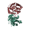
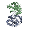
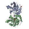
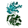
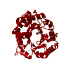
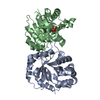
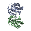
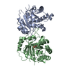
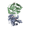
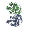
| Title | Structure of Trypanosoma brucei brucei TIM with the salt-bridge-forming residue Arg191 mutated to Ser | ||||||
 Components Components | triosephosphate isomerase, glycosomal | ||||||
 Keywords Keywords | ISOMERASE / TIM barrel / mutant / salt bridge | ||||||
| Function / homology |  Function and homology information Function and homology informationglycosome / triose-phosphate isomerase / triose-phosphate isomerase activity / glyceraldehyde-3-phosphate biosynthetic process / glycerol catabolic process / glycolytic process / gluconeogenesis / cytoplasm / cytosol Similarity search - Function | ||||||
| Biological species |  | ||||||
| Method |  X-RAY DIFFRACTION / SYNCHROTRON / MOLECULAR REPLACEMENT / Resolution: 1.65 Å X-RAY DIFFRACTION / SYNCHROTRON / MOLECULAR REPLACEMENT / Resolution: 1.65 Å | ||||||
 Authors Authors | Kursula, I. / Partanen, S. / Lambeir, A.-M. / Wierenga, R.K. | ||||||
 Citation Citation | Journal: FEBS Lett. / Year: 2002 Title: The importance of the conserved Arg191-Asp227 salt bridge of triosephosphate isomerase for folding, stability, and catalysis Authors: Kursula, I. / Partanen, S. / Lambeir, A.-M. / Wierenga, R.K. | ||||||
| History |
|
- Structure visualization
| Structure viewer | Molecule: MolmilJmol/JSmol |
|---|
- Downloads & links
-Download
| PDBx/mmCIF format | 1kv5.cif.gz | 128.4 KB | Display | PDBx/mmCIF format |
|---|---|---|---|---|
| PDB format | pdb1kv5.ent.gz | 98.3 KB | Display | PDB format |
| PDBx/mmJSON format | 1kv5.json.gz | Tree view | PDBx/mmJSON format | |
| Others |  Other downloads Other downloads |
-Validation report
| Arichive directory | https://data.pdbj.org/pub/pdb/validation_reports/kv/1kv5ftp://data.pdbj.org/pub/pdb/validation_reports/kv/1kv5 | HTTPS FTP |
|---|
-Related structure data
| Related structure data | 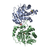 5timS S: Starting model for refinement |
|---|---|
| Similar structure data |


-Links
 PDBj
PDBj


- Assembly
| Deposited unit | 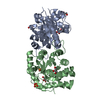
| ||||||||||
|---|---|---|---|---|---|---|---|---|---|---|---|
| 1 |
| ||||||||||
| Unit cell |
|
-Components
-Protein , 1 types, 2 molecules AB
| #1: Protein | Mass: 26795.717 Da / Num. of mol.: 2 / Mutation: R191S Source method: isolated from a genetically manipulated source Source: (gene. exp.)  |
|---|
-Non-polymers , 5 types, 787 molecules 


| #2: Chemical |  Mass: 156.031 Da / Num. of mol.: 2 Mass: 156.031 Da / Num. of mol.: 2Source method: isolated from a genetically manipulated source Formula: C2H5O6P #3: Chemical | ChemComp-DTT / |  Mass: 154.251 Da / Num. of mol.: 1 / Source method: obtained synthetically / Formula: C4H10O2S2 Mass: 154.251 Da / Num. of mol.: 1 / Source method: obtained synthetically / Formula: C4H10O2S2#4: Chemical | ChemComp-GOL /  Mass: 92.094 Da / Num. of mol.: 4 / Source method: obtained synthetically / Formula: C3H8O3 Mass: 92.094 Da / Num. of mol.: 4 / Source method: obtained synthetically / Formula: C3H8O3#5: Chemical | ChemComp-SO4 / |  Mass: 96.063 Da / Num. of mol.: 1 / Source method: obtained synthetically / Formula: SO4 Mass: 96.063 Da / Num. of mol.: 1 / Source method: obtained synthetically / Formula: SO4#6: Water | ChemComp-HOH / | Mass: 18.015 Da / Num. of mol.: 779 / Source method: isolated from a natural source / Formula: H2O |
|---|
-Experimental details
-Experiment
| Experiment | Method: X-RAY DIFFRACTION / Number of used crystals: 1 |
|---|
- Sample preparation
| Crystal | Density Matthews: 2.31 Å3/Da / Density % sol: 46.69 % | ||||||||||||||||||||||||||||||||||||||||||
|---|---|---|---|---|---|---|---|---|---|---|---|---|---|---|---|---|---|---|---|---|---|---|---|---|---|---|---|---|---|---|---|---|---|---|---|---|---|---|---|---|---|---|---|
| Crystal grow | Temperature: 295 K / Method: vapor diffusion, hanging drop / pH: 5.5 Details: ammonium sulfate, sodium chloride, citric acid, pH 5.5, VAPOR DIFFUSION, HANGING DROP at 295K | ||||||||||||||||||||||||||||||||||||||||||
| Crystal grow | *PLUS | ||||||||||||||||||||||||||||||||||||||||||
| Components of the solutions | *PLUS
|
-Data collection
| Diffraction | Mean temperature: 100 K |
|---|---|
| Diffraction source | Source: SYNCHROTRON / Site: EMBL/DESY, HAMBURG  / Beamline: X13 / Wavelength: 0.85 Å / Beamline: X13 / Wavelength: 0.85 Å |
| Detector | Type: MARRESEARCH / Detector: CCD / Date: May 27, 2001 |
| Radiation | Monochromator: Triangular monochromator / Protocol: SINGLE WAVELENGTH / Monochromatic (M) / Laue (L): M / Scattering type: x-ray |
| Radiation wavelength | Wavelength: 0.85 Å / Relative weight: 1 |
| Reflection | Resolution: 1.65→15 Å / Num. all: 115031 / Num. obs: 115031 / % possible obs: 99.9 % / Observed criterion σ(I): -3 / Redundancy: 4 % / Biso Wilson estimate: 19.5 Å2 / Rmerge(I) obs: 0.031 / Net I/σ(I): 37.7 |
| Reflection shell | Resolution: 1.65→1.68 Å / Rmerge(I) obs: 0.253 / Mean I/σ(I) obs: 5.4 / % possible all: 99.3 |
| Reflection | *PLUS Lowest resolution: 15 Å / Num. obs: 60451 / Num. measured all: 245458 |
| Reflection shell | *PLUS % possible obs: 99.3 % |
- Processing
| Software |
| ||||||||||||||||||||||||||||
|---|---|---|---|---|---|---|---|---|---|---|---|---|---|---|---|---|---|---|---|---|---|---|---|---|---|---|---|---|---|
| Refinement | Method to determine structure: MOLECULAR REPLACEMENT Starting model: PDB ENTRY 5TIM Resolution: 1.65→15 Å / Cross valid method: THROUGHOUT / σ(F): 0 / Stereochemistry target values: Engh & Huber
| ||||||||||||||||||||||||||||
| Solvent computation | Solvent model: BABINET MODEL WITH MASK PARAMETERS FOR MASK CALCULATION | ||||||||||||||||||||||||||||
| Displacement parameters | Biso mean: 15.469 Å2 | ||||||||||||||||||||||||||||
| Refinement step | Cycle: LAST / Resolution: 1.65→15 Å
| ||||||||||||||||||||||||||||
| Refine LS restraints |
| ||||||||||||||||||||||||||||
| LS refinement shell | Highest resolution: 1.65 Å / Rfactor Rfree: 0.211 / Rfactor Rwork: 0.172 / Total num. of bins used: 20 | ||||||||||||||||||||||||||||
| Refinement | *PLUS Lowest resolution: 15 Å / Rfactor obs: 0.143 / Rfactor Rfree: 0.175 / Rfactor Rwork: 0.143 | ||||||||||||||||||||||||||||
| Solvent computation | *PLUS | ||||||||||||||||||||||||||||
| Displacement parameters | *PLUS | ||||||||||||||||||||||||||||
| Refine LS restraints | *PLUS
|
