 Movie
Movie Controller

[English] 日本語
 Yorodumi
- PDB-2j24: The functional role of the conserved active site proline of trios... -
+ Open data

- Basic information
| Entry | Database: PDB / ID: 2j24 | ||||||
|---|---|---|---|---|---|---|---|
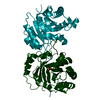
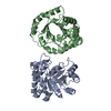
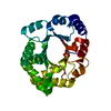
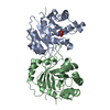
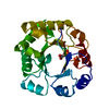
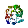
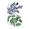
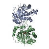
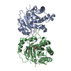
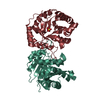
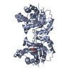
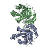
| Title | The functional role of the conserved active site proline of triosephosphate isomerase | ||||||
 Components Components | TRIOSEPHOSPHATE ISOMERASE, GLYCOSOMAL | ||||||
 Keywords Keywords | ISOMERASE / PROTEIN ENGINEERING / FATTY ACID BIOSYNTHESIS / GLUCONEOGENESIS / LIPID SYNTHESIS / PENTOSE SHUNT / POINT MUTATION / LOOP7 / GLYCOSOME / TIM-BARREL | ||||||
| Function / homology |  Function and homology information Function and homology informationglycosome / triose-phosphate isomerase / triose-phosphate isomerase activity / glyceraldehyde-3-phosphate biosynthetic process / glycerol catabolic process / glycolytic process / gluconeogenesis / cytoplasm / cytosol Similarity search - Function | ||||||
| Biological species |  | ||||||
| Method |  X-RAY DIFFRACTION / MOLECULAR REPLACEMENT / Resolution: 2.1 Å X-RAY DIFFRACTION / MOLECULAR REPLACEMENT / Resolution: 2.1 Å | ||||||
 Authors Authors | Casteleijn, M.G. / Alahuhta, M. / Groebel, K. / El-Sayed, I. / Augustyns, K. / Lambeir, A.M. / Neubauer, P. / Wierenga, R.K. | ||||||
 Citation Citation | Journal: Biochemistry / Year: 2006 Title: Functional Role of the Conserved Active Site Proline of Triosephosphate Isomerase. Authors: Casteleijn, M.G. / Alahuhta, M. / Groebel, K. / El-Sayed, I. / Augustyns, K. / Lambeir, A.M. / Neubauer, P. / Wierenga, R.K. | ||||||
| History |
| ||||||
| Remark 700 | SHEET DETERMINATION METHOD: DSSP THE SHEETS PRESENTED AS "AA" AND "BA" IN EACH CHAIN ON SHEET ... SHEET DETERMINATION METHOD: DSSP THE SHEETS PRESENTED AS "AA" AND "BA" IN EACH CHAIN ON SHEET RECORDS BELOW IS ACTUALLY AN 9-STRANDED BARREL THIS IS REPRESENTED BY A 10-STRANDED SHEET IN WHICH THE FIRST AND LAST STRANDS ARE IDENTICAL. |
- Structure visualization
| Structure viewer | Molecule: MolmilJmol/JSmol |
|---|
- Downloads & links
-Download
| PDBx/mmCIF format | 2j24.cif.gz | 115.1 KB | Display | PDBx/mmCIF format |
|---|---|---|---|---|
| PDB format | pdb2j24.ent.gz | 88.6 KB | Display | PDB format |
| PDBx/mmJSON format | 2j24.json.gz | Tree view | PDBx/mmJSON format | |
| Others |  Other downloads Other downloads |
-Validation report
| Arichive directory | https://data.pdbj.org/pub/pdb/validation_reports/j2/2j24ftp://data.pdbj.org/pub/pdb/validation_reports/j2/2j24 | HTTPS FTP |
|---|
-Related structure data
| Related structure data | 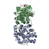 2j27C 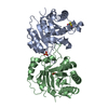 5timS S: Starting model for refinement C: citing same article ( |
|---|---|
| Similar structure data |


-Links
 PDBj
PDBj
- Assembly
| Deposited unit | 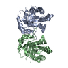
| ||||||||||||||||||
|---|---|---|---|---|---|---|---|---|---|---|---|---|---|---|---|---|---|---|---|
| 1 |
| ||||||||||||||||||
| Unit cell |
| ||||||||||||||||||
| Noncrystallographic symmetry (NCS) | NCS domain:
NCS domain segments: Component-ID: 1 / Ens-ID: 1 / Beg auth comp-ID: SER / Beg label comp-ID: SER / End auth comp-ID: GLN / End label comp-ID: GLN / Refine code: 5 / Auth seq-ID: 2 - 250 / Label seq-ID: 2 - 250
NCS oper: (Code: given Matrix: (-1, -0.000139, 0.001548), Vector: |
-Components
| #1: Protein | Mass: 26839.795 Da / Num. of mol.: 2 / Mutation: YES Source method: isolated from a genetically manipulated source Source: (gene. exp.)  #2: Water | ChemComp-HOH / |  Mass: 18.015 Da / Num. of mol.: 399 / Source method: isolated from a natural source / Formula: H2O Mass: 18.015 Da / Num. of mol.: 399 / Source method: isolated from a natural source / Formula: H2OCompound details | ENGINEERED | |
|---|
-Experimental details
-Experiment
| Experiment | Method: X-RAY DIFFRACTION / Number of used crystals: 1 |
|---|
- Sample preparation
| Crystal | Density Matthews: 2.07 Å3/Da / Density % sol: 40.54 % |
|---|---|
| Crystal grow | pH: 7 Details: WELL SOLUTION: 0.1 M TEA PH 7.0, 27% PEG 2K MME AND 0.2 M KSCN PROTEIN SOLUTION: 11 MG/ML PROTEIN, 0.02 M TRIS/HCL PH 7.0, 0.1 M NACL, 1 MM DTT, 1 MM EDTA AND 1 MM NAN3. |
-Data collection
| Diffraction | Mean temperature: 100 K |
|---|---|
| Diffraction source | Source: ROTATING ANODE / Type: ENRAF-NONIUS FR591 / Wavelength: 1.5418 |
| Detector | Type: MAR scanner 345 mm plate / Detector: IMAGE PLATE / Date: Aug 16, 2004 / Details: MONTEL MIRRORS |
| Radiation | Monochromator: MONTEL MIRRORS / Protocol: SINGLE WAVELENGTH / Monochromatic (M) / Laue (L): M / Scattering type: x-ray |
| Radiation wavelength | Wavelength: 1.5418 Å / Relative weight: 1 |
| Reflection | Resolution: 2.1→25 Å / Num. obs: 22102 / % possible obs: 94.5 % / Redundancy: 2.65 % / Biso Wilson estimate: 17.7 Å2 / Rmerge(I) obs: 0.07 / Net I/σ(I): 13.8 |
| Reflection shell | Resolution: 2.1→2.2 Å / Redundancy: 2.63 % / Rmerge(I) obs: 0.13 / Mean I/σ(I) obs: 8.67 / % possible all: 92.2 |
- Processing
| Software |
| ||||||||||||||||||||||||||||||||||||||||||||||||||||||||||||||||||||||||||||||||||||||||||||||||||||||||||||||||||||||||||||||||||||||||||||||||||||||||||||||||||||||||||||||||||||||
|---|---|---|---|---|---|---|---|---|---|---|---|---|---|---|---|---|---|---|---|---|---|---|---|---|---|---|---|---|---|---|---|---|---|---|---|---|---|---|---|---|---|---|---|---|---|---|---|---|---|---|---|---|---|---|---|---|---|---|---|---|---|---|---|---|---|---|---|---|---|---|---|---|---|---|---|---|---|---|---|---|---|---|---|---|---|---|---|---|---|---|---|---|---|---|---|---|---|---|---|---|---|---|---|---|---|---|---|---|---|---|---|---|---|---|---|---|---|---|---|---|---|---|---|---|---|---|---|---|---|---|---|---|---|---|---|---|---|---|---|---|---|---|---|---|---|---|---|---|---|---|---|---|---|---|---|---|---|---|---|---|---|---|---|---|---|---|---|---|---|---|---|---|---|---|---|---|---|---|---|---|---|---|---|
| Refinement | Method to determine structure: MOLECULAR REPLACEMENT Starting model: PDB ENTRY 5TIM Resolution: 2.1→19.21 Å / Cor.coef. Fo:Fc: 0.95 / Cor.coef. Fo:Fc free: 0.883 / SU B: 10.282 / SU ML: 0.15 / TLS residual ADP flag: UNVERIFIED / Cross valid method: THROUGHOUT / ESU R: 0.319 / ESU R Free: 0.22 / Stereochemistry target values: MAXIMUM LIKELIHOOD / Details: HYDROGENS HAVE BEEN ADDED IN THE RIDING POSITIONS.
| ||||||||||||||||||||||||||||||||||||||||||||||||||||||||||||||||||||||||||||||||||||||||||||||||||||||||||||||||||||||||||||||||||||||||||||||||||||||||||||||||||||||||||||||||||||||
| Solvent computation | Ion probe radii: 0.8 Å / Shrinkage radii: 0.8 Å / VDW probe radii: 1.2 Å / Solvent model: BABINET MODEL WITH MASK | ||||||||||||||||||||||||||||||||||||||||||||||||||||||||||||||||||||||||||||||||||||||||||||||||||||||||||||||||||||||||||||||||||||||||||||||||||||||||||||||||||||||||||||||||||||||
| Displacement parameters | Biso mean: 7.11 Å2
| ||||||||||||||||||||||||||||||||||||||||||||||||||||||||||||||||||||||||||||||||||||||||||||||||||||||||||||||||||||||||||||||||||||||||||||||||||||||||||||||||||||||||||||||||||||||
| Refinement step | Cycle: LAST / Resolution: 2.1→19.21 Å
| ||||||||||||||||||||||||||||||||||||||||||||||||||||||||||||||||||||||||||||||||||||||||||||||||||||||||||||||||||||||||||||||||||||||||||||||||||||||||||||||||||||||||||||||||||||||
| Refine LS restraints |
|