 Movie
Movie Controller

[English] 日本語
 Yorodumi
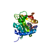
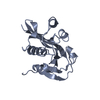
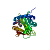
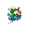
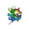
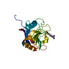
- PDB-1kuv: X-ray Crystallographic Studies of Serotonin N-acetyltransferase C... -
+ Open data

- Basic information
| Entry | Database: PDB / ID: 1kuv | ||||||
|---|---|---|---|---|---|---|---|
| Title | X-ray Crystallographic Studies of Serotonin N-acetyltransferase Catalysis and Inhibition | ||||||
 Components Components | Serotonin N-acetyltransferase | ||||||
 Keywords Keywords | TRANSFERASE / Enzyme-Inhibitor Complex / Bisubstrate Analog / Alternate Conformations | ||||||
| Function / homology |  Function and homology information Function and homology informationaralkylamine N-acetyltransferase / aralkylamine N-acetyltransferase activity / melatonin biosynthetic process / N-terminal protein amino acid acetylation / response to light stimulus / cellular response to cAMP / circadian rhythm / perinuclear region of cytoplasm Similarity search - Function | ||||||
| Biological species |  | ||||||
| Method |  X-RAY DIFFRACTION / SYNCHROTRON / MAD / Resolution: 2 Å X-RAY DIFFRACTION / SYNCHROTRON / MAD / Resolution: 2 Å | ||||||
 Authors Authors | Wolf, E. / De Angelis, J. / Khalil, E.M. / Cole, P.A. / Burley, S.K. | ||||||
 Citation Citation | Journal: J.Mol.Biol. / Year: 2002 Title: X-ray crystallographic studies of serotonin N-acetyltransferase catalysis and inhibition. Authors: Wolf, E. / De Angelis, J. / Khalil, E.M. / Cole, P.A. / Burley, S.K. | ||||||
| History |
|
- Structure visualization
| Structure viewer | Molecule: MolmilJmol/JSmol |
|---|
- Downloads & links
-Download
| PDBx/mmCIF format | 1kuv.cif.gz | 54.3 KB | Display | PDBx/mmCIF format |
|---|---|---|---|---|
| PDB format | pdb1kuv.ent.gz | 38.6 KB | Display | PDB format |
| PDBx/mmJSON format | 1kuv.json.gz | Tree view | PDBx/mmJSON format | |
| Others |  Other downloads Other downloads |
-Validation report
| Summary document | 1kuv_validation.pdf.gz | 481.2 KB | Display | wwPDB validaton report |
|---|---|---|---|---|
| Full document | 1kuv_full_validation.pdf.gz | 491.3 KB | Display | |
| Data in XML | 1kuv_validation.xml.gz | 7.3 KB | Display | |
| Data in CIF | 1kuv_validation.cif.gz | 10.4 KB | Display | |
| Arichive directory | https://data.pdbj.org/pub/pdb/validation_reports/ku/1kuvftp://data.pdbj.org/pub/pdb/validation_reports/ku/1kuv | HTTPS FTP |
-Related structure data


-Links
 PDBj
PDBj

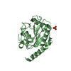
- Assembly
| Deposited unit | 
| ||||||||||
|---|---|---|---|---|---|---|---|---|---|---|---|
| 1 |
| ||||||||||
| Unit cell |
| ||||||||||
| Components on special symmetry positions |
| ||||||||||
| Details | The biological assembly is a monomer |
-Components
| #1: Protein | Mass: 23111.568 Da / Num. of mol.: 1 / Mutation: MET substituted by Se-met Source method: isolated from a genetically manipulated source Source: (gene. exp.)  References: UniProt: Q29495, aralkylamine N-acetyltransferase |
|---|---|
| #2: Chemical | ChemComp-MG /   Mass: 24.305 Da / Num. of mol.: 1 / Source method: obtained synthetically / Formula: Mg Mass: 24.305 Da / Num. of mol.: 1 / Source method: obtained synthetically / Formula: Mg |
| #3: Chemical | ChemComp-CA5 /   Mass: 1046.667 Da / Num. of mol.: 1 / Source method: obtained synthetically / Formula: C33H47BrN9O17P3S Mass: 1046.667 Da / Num. of mol.: 1 / Source method: obtained synthetically / Formula: C33H47BrN9O17P3S |
| #4: Water | ChemComp-HOH /  Mass: 18.015 Da / Num. of mol.: 167 / Source method: isolated from a natural source / Formula: H2O Mass: 18.015 Da / Num. of mol.: 167 / Source method: isolated from a natural source / Formula: H2O |
-Experimental details
-Experiment
| Experiment | Method: X-RAY DIFFRACTION / Number of used crystals: 2 |
|---|
- Sample preparation
| Crystal | Density Matthews: 1.78 Å3/Da / Density % sol: 30.81 % | ||||||||||||||||||||||||||||||||||||||||||||||||||||||||||||||||||||||
|---|---|---|---|---|---|---|---|---|---|---|---|---|---|---|---|---|---|---|---|---|---|---|---|---|---|---|---|---|---|---|---|---|---|---|---|---|---|---|---|---|---|---|---|---|---|---|---|---|---|---|---|---|---|---|---|---|---|---|---|---|---|---|---|---|---|---|---|---|---|---|---|
| Crystal grow | Temperature: 277 K / Method: vapor diffusion, hanging drop / pH: 6.5 Details: PEG 2000, MPD, ammonium sulfate, MES pH 6.5, magnesium acetate, DTT, spermidine, and lithium chloride. VAPOR DIFFUSION, HANGING DROP at 277K, temperature 277.0K | ||||||||||||||||||||||||||||||||||||||||||||||||||||||||||||||||||||||
| Crystal grow | *PLUS Temperature: 4 ℃ / Details: used microseeding | ||||||||||||||||||||||||||||||||||||||||||||||||||||||||||||||||||||||
| Components of the solutions | *PLUS
|
-Data collection
| Diffraction |
| ||||||||||||||||||
|---|---|---|---|---|---|---|---|---|---|---|---|---|---|---|---|---|---|---|---|
| Diffraction source |
| ||||||||||||||||||
| Detector |
| ||||||||||||||||||
| Radiation |
| ||||||||||||||||||
| Radiation wavelength |
| ||||||||||||||||||
| Reflection | Resolution: 2→25 Å / Num. all: 11494 / Num. obs: 10517 / % possible obs: 91.5 % / Observed criterion σ(I): -3 / Redundancy: 8.3 % / Rmerge(I) obs: 0.089 / Rsym value: 0.089 / Net I/σ(I): 16.3 | ||||||||||||||||||
| Reflection shell | Resolution: 2→2.07 Å / Redundancy: 5 % / Rmerge(I) obs: 0.123 / Mean I/σ(I) obs: 9.6 / Num. unique all: 1148 / Rsym value: 0.123 / % possible all: 79.5 | ||||||||||||||||||
| Reflection | *PLUS Highest resolution: 2 Å / Lowest resolution: 25 Å / % possible obs: 92 % / Num. measured all: 86780 | ||||||||||||||||||
| Reflection shell | *PLUS % possible obs: 80 % |

- Processing
| Software |
| |||||||||||||||||||||||||||
|---|---|---|---|---|---|---|---|---|---|---|---|---|---|---|---|---|---|---|---|---|---|---|---|---|---|---|---|---|
| Refinement | Method to determine structure: MAD / Resolution: 2→25 Å / Cross valid method: THROUGHOUT / σ(F): 0 / σ(I): 0 / Stereochemistry target values: Engh & Huber Details: Structure was solved based on MAD data collected in the vicinity of the bromine and selenium edges. Ligand density was identified by difference fourier. The final model (protein and ligand) ...Details: Structure was solved based on MAD data collected in the vicinity of the bromine and selenium edges. Ligand density was identified by difference fourier. The final model (protein and ligand) was refined at 2.0 Angstrom
| |||||||||||||||||||||||||||
| Displacement parameters |
| |||||||||||||||||||||||||||
| Refinement step | Cycle: LAST / Resolution: 2→25 Å
| |||||||||||||||||||||||||||
| Refine LS restraints |
| |||||||||||||||||||||||||||
| Refinement | *PLUS Lowest resolution: 25 Å / Num. reflection obs: 10057 / % reflection Rfree: 10 % / Rfactor obs: 0.203 / Rfactor Rfree: 0.25 | |||||||||||||||||||||||||||
| Solvent computation | *PLUS | |||||||||||||||||||||||||||
| Displacement parameters | *PLUS |
