 Movie
Movie Controller

[English] 日本語
 Yorodumi
- PDB-1l0c: Investigation of the Roles of Catalytic Residues in Serotonin N-A... -
+ Open data

- Basic information
| Entry | Database: PDB / ID: 1l0c | ||||||
|---|---|---|---|---|---|---|---|
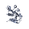
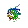
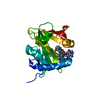
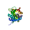
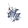
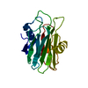
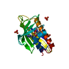
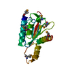
| Title | Investigation of the Roles of Catalytic Residues in Serotonin N-Acetyltransferase | ||||||
 Components Components | Serotonin N-acetyltransferase | ||||||
 Keywords Keywords | TRANSFERASE / Enzyme-inhibitor complex / bisubstrate analog / alternate conformations | ||||||
| Function / homology |  Function and homology information Function and homology informationmelatonin biosynthetic process / aralkylamine N-acetyltransferase / N-terminal protein amino acid acetylation / aralkylamine N-acetyltransferase activity / response to light stimulus / cellular response to cAMP / circadian rhythm / perinuclear region of cytoplasm Similarity search - Function | ||||||
| Biological species |  | ||||||
| Method |  X-RAY DIFFRACTION / SYNCHROTRON / MOLECULAR REPLACEMENT / Resolution: 2.3 Å X-RAY DIFFRACTION / SYNCHROTRON / MOLECULAR REPLACEMENT / Resolution: 2.3 Å | ||||||
 Authors Authors | Scheibner, K.A. / De Angelis, J. / Burley, S.K. / Cole, P.A. | ||||||
 Citation Citation | Journal: J.Biol.Chem. / Year: 2002 Title: Investigation of the roles of catalytic residues in serotonin N-acetyltransferase. Authors: Scheibner, K.A. / De Angelis, J. / Burley, S.K. / Cole, P.A. | ||||||
| History |
|
- Structure visualization
| Structure viewer | Molecule: MolmilJmol/JSmol |
|---|
- Downloads & links
-Download
| PDBx/mmCIF format | 1l0c.cif.gz | 50.7 KB | Display | PDBx/mmCIF format |
|---|---|---|---|---|
| PDB format | pdb1l0c.ent.gz | 34.7 KB | Display | PDB format |
| PDBx/mmJSON format | 1l0c.json.gz | Tree view | PDBx/mmJSON format | |
| Others |  Other downloads Other downloads |
-Validation report
| Arichive directory | https://data.pdbj.org/pub/pdb/validation_reports/l0/1l0cftp://data.pdbj.org/pub/pdb/validation_reports/l0/1l0c | HTTPS FTP |
|---|
-Related structure data
| Related structure data | 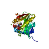 1kuvS S: Starting model for refinement |
|---|---|
| Similar structure data |


-Links
 PDBj
PDBj

- Assembly
| Deposited unit | 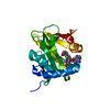
| ||||||||
|---|---|---|---|---|---|---|---|---|---|
| 1 |
| ||||||||
| Unit cell |
| ||||||||
| Details | The biological assembly is a monomer. |
-Components
| #1: Protein | Mass: 23095.568 Da / Num. of mol.: 1 / Mutation: Y168F Source method: isolated from a genetically manipulated source Source: (gene. exp.)  References: UniProt: Q29495, aralkylamine N-acetyltransferase |
|---|---|
| #2: Chemical | ChemComp-COT /   Mass: 967.771 Da / Num. of mol.: 1 / Source method: obtained synthetically / Formula: C33H48N9O17P3S Mass: 967.771 Da / Num. of mol.: 1 / Source method: obtained synthetically / Formula: C33H48N9O17P3S |
| #3: Water | ChemComp-HOH /  Mass: 18.015 Da / Num. of mol.: 90 / Source method: isolated from a natural source / Formula: H2O Mass: 18.015 Da / Num. of mol.: 90 / Source method: isolated from a natural source / Formula: H2O |
-Experimental details
-Experiment
| Experiment | Method: X-RAY DIFFRACTION / Number of used crystals: 1 |
|---|
- Sample preparation
| Crystal grow | Temperature: 277 K / Method: vapor diffusion, hanging drop / pH: 6.5 Details: PEG 2000, MPD, MES pH 6.5, ammonium acetate, magnesium acetate, lithium chloride, spermidine, DTT, VAPOR DIFFUSION, HANGING DROP, temperature 277K | ||||||||||||||||||||||||||||||||||||||||||||||||||||||||||||||||||||||
|---|---|---|---|---|---|---|---|---|---|---|---|---|---|---|---|---|---|---|---|---|---|---|---|---|---|---|---|---|---|---|---|---|---|---|---|---|---|---|---|---|---|---|---|---|---|---|---|---|---|---|---|---|---|---|---|---|---|---|---|---|---|---|---|---|---|---|---|---|---|---|---|
| Crystal grow | *PLUS Temperature: 4 ℃Details: Wolf, E., (2002) J. Mol. Biol., 317, 215., used microseeding | ||||||||||||||||||||||||||||||||||||||||||||||||||||||||||||||||||||||
| Components of the solutions | *PLUS
|
-Data collection
| Diffraction | Mean temperature: 100 K |
|---|---|
| Diffraction source | Source: SYNCHROTRON / Site: NSLS  / Beamline: X4A / Wavelength: 0.979 Å / Beamline: X4A / Wavelength: 0.979 Å |
| Detector | Type: ADSC QUANTUM 4 / Detector: CCD / Date: Apr 13, 2000 |
| Radiation | Monochromator: Si 111 / Protocol: SINGLE WAVELENGTH / Monochromatic (M) / Laue (L): M / Scattering type: x-ray |
| Radiation wavelength | Wavelength: 0.979 Å / Relative weight: 1 |
| Reflection | Resolution: 2.3→20 Å / Num. all: 7822 / Num. obs: 7188 / % possible obs: 91.9 % / Observed criterion σ(F): -3 / Observed criterion σ(I): -3 / Redundancy: 4 % / Rmerge(I) obs: 0.037 / Rsym value: 0.037 / Net I/σ(I): 25.2 |
| Reflection shell | Resolution: 2.3→2.35 Å / Rmerge(I) obs: 0.092 / Mean I/σ(I) obs: 14.1 / Num. unique all: 497 / Rsym value: 0.092 / % possible all: 93 |
| Reflection | *PLUS Lowest resolution: 20 Å / Num. measured all: 75910 / Rmerge(I) obs: 0.037 |
| Reflection shell | *PLUS % possible obs: 93 % / Rmerge(I) obs: 0.092 |
- Processing
| Software |
| |||||||||||||||||||||
|---|---|---|---|---|---|---|---|---|---|---|---|---|---|---|---|---|---|---|---|---|---|---|
| Refinement | Method to determine structure: MOLECULAR REPLACEMENT Starting model: PDB ENTRY 1KUV Resolution: 2.3→20 Å / Cross valid method: THROUGHOUT / σ(F): 0 / σ(I): 0 / Stereochemistry target values: Engh & Huber Details: STRUCTURE WAS REFINED WITH WATERS AND LIGAND WAS MODELED BASED ON DIFFERENCE FOURIER ELECTRON DENSITY. LIGAND WAS INCLUDED IN FINAL REFINEMENT.
| |||||||||||||||||||||
| Displacement parameters |
| |||||||||||||||||||||
| Refinement step | Cycle: LAST / Resolution: 2.3→20 Å
| |||||||||||||||||||||
| Refine LS restraints |
| |||||||||||||||||||||
| Refinement | *PLUS Lowest resolution: 20 Å / Rfactor obs: 0.1877 / Rfactor Rfree: 0.246 / Rfactor Rwork: 0.193 | |||||||||||||||||||||
| Solvent computation | *PLUS | |||||||||||||||||||||
| Displacement parameters | *PLUS |
