 Movie
Movie Controller

[English] 日本語
 Yorodumi
- PDB-1cjw: SEROTONIN N-ACETYLTRANSFERASE COMPLEXED WITH A BISUBSTRATE ANALOG -
+ Open data

- Basic information
| Entry | Database: PDB / ID: 1cjw | ||||||
|---|---|---|---|---|---|---|---|
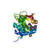
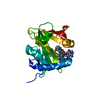
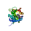
| Title | SEROTONIN N-ACETYLTRANSFERASE COMPLEXED WITH A BISUBSTRATE ANALOG | ||||||
 Components Components | PROTEIN (SEROTONIN N-ACETYLTRANSFERASE) | ||||||
 Keywords Keywords | TRANSFERASE / N-ACETYL TRANSFERASE | ||||||
| Function / homology |  Function and homology information Function and homology informationmelatonin biosynthetic process / aralkylamine N-acetyltransferase / N-terminal protein amino acid acetylation / aralkylamine N-acetyltransferase activity / response to light stimulus / cellular response to cAMP / circadian rhythm / perinuclear region of cytoplasm Similarity search - Function | ||||||
| Biological species |  | ||||||
| Method |  X-RAY DIFFRACTION / MOLECULAR REPLACEMENT / Resolution: 1.8 Å X-RAY DIFFRACTION / MOLECULAR REPLACEMENT / Resolution: 1.8 Å | ||||||
 Authors Authors | Hickman, A.B. / Namboodiri, M.A.A. / Klein, D.C. / Dyda, F. | ||||||
 Citation Citation | Journal: Cell(Cambridge,Mass.) / Year: 1999 Title: The structural basis of ordered substrate binding by serotonin N-acetyltransferase: enzyme complex at 1.8 A resolution with a bisubstrate analog. Authors: Hickman, A.B. / Namboodiri, M.A. / Klein, D.C. / Dyda, F. | ||||||
| History |
|
- Structure visualization
| Structure viewer | Molecule: MolmilJmol/JSmol |
|---|
- Downloads & links
-Download
| PDBx/mmCIF format | 1cjw.cif.gz | 55 KB | Display | PDBx/mmCIF format |
|---|---|---|---|---|
| PDB format | pdb1cjw.ent.gz | 37.4 KB | Display | PDB format |
| PDBx/mmJSON format | 1cjw.json.gz | Tree view | PDBx/mmJSON format | |
| Others |  Other downloads Other downloads |
-Validation report
| Arichive directory | https://data.pdbj.org/pub/pdb/validation_reports/cj/1cjwftp://data.pdbj.org/pub/pdb/validation_reports/cj/1cjw | HTTPS FTP |
|---|
-Related structure data
| Related structure data |  1b6bS S: Starting model for refinement |
|---|---|
| Similar structure data |


-Links
 PDBj
PDBj

- Assembly
| Deposited unit | 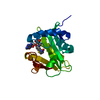
| ||||||||
|---|---|---|---|---|---|---|---|---|---|
| 1 |
| ||||||||
| Unit cell |
|
-Components
| #1: Protein | Mass: 18634.381 Da / Num. of mol.: 1 Source method: isolated from a genetically manipulated source Source: (gene. exp.)  |
|---|---|
| #2: Chemical | ChemComp-COT /   Mass: 967.771 Da / Num. of mol.: 1 / Source method: obtained synthetically / Formula: C33H48N9O17P3S Mass: 967.771 Da / Num. of mol.: 1 / Source method: obtained synthetically / Formula: C33H48N9O17P3S |
| #3: Water | ChemComp-HOH /  Mass: 18.015 Da / Num. of mol.: 261 / Source method: isolated from a natural source / Formula: H2O Mass: 18.015 Da / Num. of mol.: 261 / Source method: isolated from a natural source / Formula: H2O |
-Experimental details
-Experiment
| Experiment | Method: X-RAY DIFFRACTION / Number of used crystals: 1 |
|---|
- Sample preparation
| Crystal | Density Matthews: 2.21 Å3/Da / Density % sol: 44.28 % | ||||||||||||||||||||
|---|---|---|---|---|---|---|---|---|---|---|---|---|---|---|---|---|---|---|---|---|---|
| Crystal grow | pH: 6.5 / Details: pH 6.5 | ||||||||||||||||||||
| Crystal | *PLUS Density % sol: 34.9 % | ||||||||||||||||||||
| Crystal grow | *PLUS Temperature: 4 ℃ / Method: vapor diffusion, hanging drop | ||||||||||||||||||||
| Components of the solutions | *PLUS
|
-Data collection
| Diffraction | Mean temperature: 100 K |
|---|---|
| Diffraction source | Source: ROTATING ANODE / Type: RIGAKU RUH3R / Wavelength: 1.5418 |
| Detector | Type: RIGAKU RAXIS IV / Detector: IMAGE PLATE / Date: Dec 1, 1998 / Details: MIRRORS |
| Radiation | Monochromator: NI FILTER / Protocol: SINGLE WAVELENGTH / Monochromatic (M) / Laue (L): M / Scattering type: x-ray |
| Radiation wavelength | Wavelength: 1.5418 Å / Relative weight: 1 |
| Reflection | Resolution: 1.8→30 Å / Num. obs: 15197 / % possible obs: 97 % / Observed criterion σ(I): 0 / Redundancy: 6.3 % / Rsym value: 0.041 / Net I/σ(I): 18 |
| Reflection shell | Resolution: 1.8→1.85 Å / Redundancy: 3.5 % / Mean I/σ(I) obs: 11 / Rsym value: 0.121 / % possible all: 97 |
| Reflection | *PLUS Num. measured all: 96003 / Rmerge(I) obs: 0.041 |
| Reflection shell | *PLUS % possible obs: 97.4 % / Rmerge(I) obs: 0.121 |
- Processing
| Software |
| ||||||||||||||||||||||||||||||||||||||||||||||||||||||||||||
|---|---|---|---|---|---|---|---|---|---|---|---|---|---|---|---|---|---|---|---|---|---|---|---|---|---|---|---|---|---|---|---|---|---|---|---|---|---|---|---|---|---|---|---|---|---|---|---|---|---|---|---|---|---|---|---|---|---|---|---|---|---|
| Refinement | Method to determine structure: MOLECULAR REPLACEMENT Starting model: 1B6B Resolution: 1.8→30 Å / Rfactor Rfree error: 0.008 / Cross valid method: THROUGHOUT / σ(F): 2
| ||||||||||||||||||||||||||||||||||||||||||||||||||||||||||||
| Refinement step | Cycle: LAST / Resolution: 1.8→30 Å
| ||||||||||||||||||||||||||||||||||||||||||||||||||||||||||||
| Refine LS restraints |
| ||||||||||||||||||||||||||||||||||||||||||||||||||||||||||||
| LS refinement shell | Resolution: 1.8→1.88 Å / Rfactor Rfree error: 0.034 / Total num. of bins used: 8
| ||||||||||||||||||||||||||||||||||||||||||||||||||||||||||||
| Xplor file | Serial no: 1 / Param file: PARHCSDX.PRO / Topol file: TOPHCSDX.PRO | ||||||||||||||||||||||||||||||||||||||||||||||||||||||||||||
| Software | *PLUS Name: X-PLOR / Version: 3.1 / Classification: refinement | ||||||||||||||||||||||||||||||||||||||||||||||||||||||||||||
| Refinement | *PLUS Rfactor obs: 0.181 / Rfactor Rfree: 0.231 | ||||||||||||||||||||||||||||||||||||||||||||||||||||||||||||
| Solvent computation | *PLUS | ||||||||||||||||||||||||||||||||||||||||||||||||||||||||||||
| Displacement parameters | *PLUS |
