 Movie
Movie Controller

[English] 日本語
 Yorodumi
- PDB-1b6b: MELATONIN BIOSYNTHESIS: THE STRUCTURE OF SEROTONIN N-ACETYLTRANSF... -
+ Open data

- Basic information
| Entry | Database: PDB / ID: 1b6b | ||||||
|---|---|---|---|---|---|---|---|
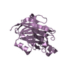
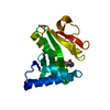
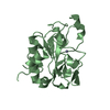
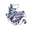
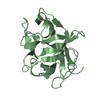
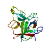
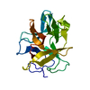
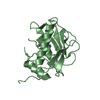
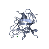
| Title | MELATONIN BIOSYNTHESIS: THE STRUCTURE OF SEROTONIN N-ACETYLTRANSFERASE AT 2.5 A RESOLUTION SUGGESTS A CATALYTIC MECHANISM | ||||||
 Components Components | PROTEIN (ARYLALKYLAMINE N-ACETYLTRANSFERASE) | ||||||
 Keywords Keywords | TRANSFERASE / ACETYLTRANSFERASE | ||||||
| Function / homology |  Function and homology information Function and homology informationmelatonin biosynthetic process / aralkylamine N-acetyltransferase / aralkylamine N-acetyltransferase activity / N-terminal protein amino acid acetylation / response to light stimulus / cellular response to cAMP / circadian rhythm / perinuclear region of cytoplasm Similarity search - Function | ||||||
| Biological species |  | ||||||
| Method |  X-RAY DIFFRACTION / SYNCHROTRON / MAD / Resolution: 2.5 Å X-RAY DIFFRACTION / SYNCHROTRON / MAD / Resolution: 2.5 Å | ||||||
 Authors Authors | Hickman, A.B. / Klein, D.C. / Dyda, F. | ||||||
 Citation Citation | Journal: Mol.Cell / Year: 1999 Title: Melatonin biosynthesis: the structure of serotonin N-acetyltransferase at 2.5 A resolution suggests a catalytic mechanism. Authors: Hickman, A.B. / Klein, D.C. / Dyda, F. | ||||||
| History |
|
- Structure visualization
| Structure viewer | Molecule: MolmilJmol/JSmol |
|---|
- Downloads & links
-Download
| PDBx/mmCIF format | 1b6b.cif.gz | 76.4 KB | Display | PDBx/mmCIF format |
|---|---|---|---|---|
| PDB format | pdb1b6b.ent.gz | 57 KB | Display | PDB format |
| PDBx/mmJSON format | 1b6b.json.gz | Tree view | PDBx/mmJSON format | |
| Others |  Other downloads Other downloads |
-Validation report
| Arichive directory | https://data.pdbj.org/pub/pdb/validation_reports/b6/1b6bftp://data.pdbj.org/pub/pdb/validation_reports/b6/1b6b | HTTPS FTP |
|---|
-Related structure data
| Similar structure data |
|---|

-Links
 PDBj
PDBj

- Assembly
| Deposited unit | 
| ||||||||
|---|---|---|---|---|---|---|---|---|---|
| 1 | 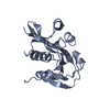
| ||||||||
| 2 | 
| ||||||||
| Unit cell |
| ||||||||
| Noncrystallographic symmetry (NCS) | NCS oper: (Code: given Matrix: (-0.8237, -0.5629, -0.0681), Vector: |
-Components
| #1: Protein | Mass: 19556.477 Da / Num. of mol.: 2 Source method: isolated from a genetically manipulated source Source: (gene. exp.)  References: UniProt: Q29495, aralkylamine N-acetyltransferase #2: Water | ChemComp-HOH / |  Mass: 18.015 Da / Num. of mol.: 132 / Source method: isolated from a natural source / Formula: H2O Mass: 18.015 Da / Num. of mol.: 132 / Source method: isolated from a natural source / Formula: H2O |
|---|
-Experimental details
-Experiment
| Experiment | Method: X-RAY DIFFRACTION / Number of used crystals: 1 |
|---|
- Sample preparation
| Crystal | Density Matthews: 2.46 Å3/Da / Density % sol: 51 % | ||||||||||||||||||||||||||||||||||||
|---|---|---|---|---|---|---|---|---|---|---|---|---|---|---|---|---|---|---|---|---|---|---|---|---|---|---|---|---|---|---|---|---|---|---|---|---|---|
| Crystal grow | pH: 6.5 / Details: pH 6.5 | ||||||||||||||||||||||||||||||||||||
| Crystal | *PLUS Density % sol: 49 % | ||||||||||||||||||||||||||||||||||||
| Crystal grow | *PLUS Method: microdialysis | ||||||||||||||||||||||||||||||||||||
| Components of the solutions | *PLUS
|
-Data collection
| Diffraction | Mean temperature: 95 K | ||||||||||||
|---|---|---|---|---|---|---|---|---|---|---|---|---|---|
| Diffraction source | Source: SYNCHROTRON / Site: NSLS  / Beamline: X9B / Wavelength: 0.97935, 0.97900, 0.96860 / Beamline: X9B / Wavelength: 0.97935, 0.97900, 0.96860 | ||||||||||||
| Detector | Type: MARRESEARCH / Detector: IMAGE PLATE / Date: Jun 15, 1998 / Details: MIRROR | ||||||||||||
| Radiation | Monochromator: SI111 / Protocol: MAD / Monochromatic (M) / Laue (L): M / Scattering type: x-ray | ||||||||||||
| Radiation wavelength |
| ||||||||||||
| Reflection | Resolution: 3→20 Å / Num. obs: 6476 / % possible obs: 85.4 % / Observed criterion σ(I): 0 / Redundancy: 5 % / Rsym value: 6.3 / Net I/σ(I): 14.5 | ||||||||||||
| Reflection shell | Resolution: 3→3.11 Å / Redundancy: 3 % / Mean I/σ(I) obs: 10.5 / Rsym value: 13.2 / % possible all: 93.2 | ||||||||||||
| Reflection | *PLUS Num. measured all: 39960 / Rmerge(I) obs: 0.063 | ||||||||||||
| Reflection shell | *PLUS % possible obs: 93.2 % / Rmerge(I) obs: 0.132 |
- Processing
| Software |
| ||||||||||||||||||||||||||||||||||||||||||||||||||||||||||||
|---|---|---|---|---|---|---|---|---|---|---|---|---|---|---|---|---|---|---|---|---|---|---|---|---|---|---|---|---|---|---|---|---|---|---|---|---|---|---|---|---|---|---|---|---|---|---|---|---|---|---|---|---|---|---|---|---|---|---|---|---|---|
| Refinement | Method to determine structure: MAD / Resolution: 2.5→30 Å / Cross valid method: THROUGHOUT / σ(F): 2 Details: IN STRUCTURE DETERMINATION, A TRUNCATED CONSTRUCT WAS USED BETWEEN RESIDUES 27 - 201
| ||||||||||||||||||||||||||||||||||||||||||||||||||||||||||||
| Refinement step | Cycle: LAST / Resolution: 2.5→30 Å
| ||||||||||||||||||||||||||||||||||||||||||||||||||||||||||||
| Refine LS restraints |
| ||||||||||||||||||||||||||||||||||||||||||||||||||||||||||||
| Xplor file | Serial no: 1 / Param file: PARHCSDX.PRO / Topol file: TOPHCSDX.PRO | ||||||||||||||||||||||||||||||||||||||||||||||||||||||||||||
| Software | *PLUS Name: X-PLOR / Version: 3.1 / Classification: refinement | ||||||||||||||||||||||||||||||||||||||||||||||||||||||||||||
| Refine LS restraints | *PLUS
|
